Архів офтальмології та щелепно-лицевої хірургії України Том 1, №1, 2024
Вернуться к номеру
Аналіз морфофункціональних змін сітківки після вітректомії у пацієнтів з регматогенним відшаруванням
Авторы: Іванченко А.Ю., Безкоровайна І.М.
Полтавський державний медичний університет, м. Полтава, Україна
Рубрики: Офтальмология
Разделы: Клинические исследования
Версия для печати
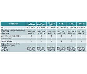
Актуальність. Отримання максимально високих функціональних даних після лікування регматогенного відшарування сітківки (РВС) є важливою темою протягом останніх кількох десятиліть. Розширення знань стосовно морфологічних змін макулярної ділянки та хоріоретинального кровотоку може сприяти вирішенню низки питань стосовно цієї проблеми. Мета. Вивчити взаємозв’язок гостроти зору (ГЗ) зі змінами мікрокровотоку макулярної ділянки сітківки після вітректомії з приводу регматогенного відшарування сітківки. Матеріали та методи. У дослідженні взяли участь 35 пацієнтів із первинним РВС. Усім було проведено задню субтотальну вітректомію з ендовітреальною тампонадою силіконовою олією та виведенням через 1 місяць. Аналіз змін макулярної ділянки виконували на оптичному когерентному томографі. Внутрішньоочний кровотік досліджували за допомогою оптичної когерентної томографії з функцією ангіографії. Результати. Отримані дані продемонстрували залежність рівня ГЗ від стану мікроструктури і хоріоретинального кровотоку макулярної ділянки на завершальному етапі силіконової тампонади при ендовітреальній хірургії РВС. Висновки. Основною причиною низької ГЗ є структурні зміни нейроепітелію в макулі, дезорганізація лінії еліпсоїдної зони, дефекти зовнішньої межової мембрани та внутрішньої межової мембрани. Виявлено взаємозв’язок між ступенем зниження ГЗ, морфологічними змінами в макулі та тяжкістю порушення хоріоретинального кровотоку в макулі.
Background. Obtaining the highest possible functional data after treatment for rhegmatogenous retinal detachment (RRD) has been an important topic over the past few decades. Expanding knowledge of the morphological changes in the macular area and chorioretinal blood flow may help resolve a number of issues related to this problem. Objective: to study the relationship between visual acuity (VA) and changes in macular microcirculation after vitrectomy for rhegmatogenous retinal detachment. Materials and methods. The study involved 35 patients with primary RRD. All of them underwent posterior subtotal vitrectomy with endovitreal tamponade using silicone oil and withdrawal after 1 month. Changes in the macular area were analysed by optical coherence tomography. The intraocular blood flow was studied using optical coherence tomography with angiography function. Results. The data obtained demonstrated the dependence of the level of VA on the state of the microstructure and chorioretinal blood flow in the macular area at the final stage of silicone tamponade during endovitreal surgery for RRD. Conclusions. The main cause of low VA is structural changes in the neuroepithelium in the macula, disorganisation of the ellipsoid zone line, defects in the external and internal limiting membrane. The correlation was found between the degree of VA reduction, morphological changes in the macula and the severity of chorioretinal blood flow disorders in the macula.
регматогенне відшарування сітківки; ОКТ-ангіографія; макула
rhegmatogenous retinal detachment; optical coherence tomography angiography; macula
Для ознакомления с полным содержанием статьи необходимо оформить подписку на журнал.
- Muni RH, Darabad MN, Oquendo PL et al. Outer retinal corrugations in rhegmatogenous retinal detachment: the retinal pigment epithelium-photoreceptor dysregulation theory. Am J Ophthalmol. 2023;245:14-24. doi: 10.1016/j.ajo.2022.08.019.
- Ambiya V, Rani P et al. Outcomes of recurrent retinal detachment surgery following pars plana vitrectomy for rhegmatogenous retinal detachment, Seminars in Ophthalmology. 2018;33(5):657-663. https://doi.org/10.1080/08820538.2017.1395893.
- Grabowska A, Neffendorf J, Yorston D, Williamson T. Urgency of retinal detachment repair: is it time to re-think our priorities? Eye (Lond). 2021;35:1035-1036.
- Noda H, Kimura S, Morizane Y, Toshima S et al. Relationship between preoperative foveal microstructure and visual acuity in macula-off rhegmatogenous retinal detachment: imaging analysis by swept-source optical coherence tomography. Retina. 2020;40(10):1873-1880. doi: 10.1097/IAE.0000000000002687.
- Melo IM, Bansal A, Lee WW et al. Bacillary layer detachment and associated abnormalities in rhegmatogenous retinal detachment. Retina. 2023;43(4):670-678. doi: 10.1097/IAE.0000000000003696.
- Hostovsky A, Trussart R, AlAli A et al. Pre-operative optical coherence tomography findings in macula-off retinal detachments and visual outcome. Eye (Lond). 2021;35(12):3285-3291. doi: 10.1038/s41433-021-01399-z.
- Terauchi G, Shinoda K, Matsumoto C, Watanabe E, Matsumoto H, Mizota A. Recovery of photoreceptor inner and outer segment layer thickness after reattachment of rhegmatogenous retinal detachment. Br. J. Ophthalmol. 2015;99(10):1323-1327. doi: 10.1136/bjophthalmol-2014-306252.
- Inan S, Polat O, Ozcan S, Inan U. Comparison of long-term automated retinal layer segmentation analysis of the macula between silicone oil and gas tamponade after vitrectomy for rhegmatogenous retinal detachment. Ophthalmic Res. 2020;63(6):524-532. https://doi.org/–10.1159/000506382.
- Hostovsky A, Trussart R, AlAli A, Kertes PJ, Eng KT. Pre-–operative optical coherence tomography findings in macula-off retinal detachments and visual outcome. Eye. 2021;35(12):3285-3291. doi: 10.1038/s41433-021-01399-z.
- Bonfiglio V, Ortisi E, Scollo D, Reibaldi M, Russo A, Pizzo A et al. Vascular changes after vitrectomy for rhegmatogenous retinal detachment: Optical coherence tomography angiography study. Acta Ophthalmol. 2020;98(5):e563-e569. doi: 10.1111/aos.14315.

