Журнал «Травма» Том 24, №2, 2023
Вернуться к номеру
Вплив низькочастотної вібрації на відновлення обсягу рухів колінного суглоба лабораторних тварин після іммобілізації (експериментальне дослідження)
Авторы: Тяжелов О.А. (1), Карпінська О.Д. (1), Карпінський М.Ю. (1), Нікольченко О.А. (1), Фіщенко В.О. (2), Хасавнех Айхам Адлі Мохаммад (2)
(1) — ДУ «Інститут патології хребта та суглобів ім. проф. М.І. Ситенка НАМН України», м. Харків, Україна
(2) — Вінницкий національний медичний університет ім. М.І. Пирогова МОЗ України, м. Вінниця, Україна
Рубрики: Травматология и ортопедия
Разделы: Клинические исследования
Версия для печати
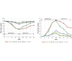
Актуальність. Термін «суглобові контрактури» використовують для опису втрати пасивного діапазону рухів діартрозних суглобів, найбільш поширеного та рухомого типу суглобів. Вимірювання пасивного чи активного діапазону рухів у суглобі з контрактурою є ключем до оцінки важливості контрактур суглобів. Мета дослідження: визначити вплив іммобілізації на розвиток обмеження рухів у колінному суглобі лабораторних тварин (щурів) та оцінити можливість відновлення рухливості у разі використання низькочастотної вібрації в процесі іммобілізації та після її завершення. Матеріали та методи. Експериментальне дослідження проведене на 30 нелінійних білих щурах-самцях віком 6 місяців. Іммобілізацію тазової кінцівки виконували під кутом 140° у колінному суглобі. Тварини випадковим чином були поділені на 3 групи: І — іммобілізація та вільне утримання після іммобілізації, ІІ — іммобілізація та вібраційна розробка суглоба після іммобілізації, ІІІ — іммобілізація та вібраційна розробка суглоба під час і після іммобілізації. Вібраційну розробку іммобілізованого колінного суглоба проводили щоденно в режимі 20 Гц з амплітудою 1,5 мм тривалістю 10 хвилин. Визначали обсяг рухів та реальну контрактуру як різницю між виміряним обсягом рухів та обсягом рухів перед початком експерименту для кожної тварини індивідуально. Результати. Було виявлено, що стрімке наростання обмеження рухів відбувається починаючи з 2-го тижня іммобілізації. Зменшення обсягу рухів у щурів І та ІІ груп в умовах іммобілізації відбувалося однаково. Після завершення іммобілізації в І групі спостерігали повільне зростання обсягу рухів, в ІІ групі зростання було майже лінійним, і через 4 тижні показник був близький до норми. У ІІІ групі обмеження обсягу рухів після іммобілізації було значуще меншим, отже, і відновлення відбулося вже через 2 тижні після зняття іммобілізаційної пов’язки. Іммобілізація колінного суглоба у щурів І та ІІ груп спричинила контрактуру в 60°, тоді як у ІІІ групі обмеження не перевищували 25°. І відповідно, відновлення в групах з вібраційною розробкою було стрімким, в ІІІ групі досягнуто повне відновлення, в ІІ групі — відновлення до 5° залишкової контрактури. В І групі спостерігаємо залишкову контрактуру у майже 35°, що більше, ніж сформована іммобілізаційна контрактура в ІІІ групі. Висновки. Низькочастотна вібрація дозволяє зменшити вплив іммобілізації і значно прискорити відновлення рухливості (обсягу рухів) суглоба після її завершення. За неможливості проведення вібротерапії в період іммобілізації її слід починати якомога раніше після іммобілізації. На сьогодні існує мало досліджень щодо впливу низькочастотної вібрації на розвиток іммобілізаційних контрактур та їх лікування. Отримані дані потребують подальших досліджень із більш тривалими термінами іммобілізації та стосовно варіантів іммобілізації і режимів вібраційного впливу на суглоби.
Background. The term “joint contractures” is used to describe the loss of passive range of motion of diarthrosis joints, the most common and mobile type of a joint. Measuring passive or active range of motion in a joint with contracture is key to assessing the severity of joint contractures. The purpose of the study: to determine the impact of immobilization on the development of movement limitation in the knee joint of laboratory animals (rats) and to evaluate the possibility of restoring mobility in case of using low-frequency vibration during and after immobilization. Materials and methods. The experimental study was conducted on 30 non-linear white male rats aged 6 months. Immobilization of the pelvic limb was performed at an angle of 140° in the knee joint. The animals were randomly divided into 3 groups: I — immobilization and free restraint after immobilization, II — immobilization and vibration development of the joint after immobilization, III — immobilization and vibration development of the joint during and after immobilization. Vibration development of the immobilized knee joint was performed daily in the mode of 20 Hz with an amplitude of 1.5 mm and a duration of 10 minutes. The range of motion and real contracture were determined as the difference between the measured range of motion and the range of motion before the start of the experiment for each animal individually. Results. It was found that a rapid increase in movement limitation occurs starting from the 2nd week of immobilization. A decrease in the range of motion in rats of the groups I and II under conditions of immobilization occurred the same way. After the end of immobilization, a slow increase in the range of motion was observed in the group I; in the group II, the growth was almost linear and after 4 weeks, the indicator was close to the norm. In the group III, the limitation of the range of motion after immobilization was significantly less; therefore, accordingly, recovery took place already 2 weeks after the removal of the immobilization bandage. Immobilization of the knee joint in rats of groups I and II caused a contracture of 60°, while in the group III, the restrictions did not exceed 25°. And, accordingly, the recovery in the groups with vibration development was rapid; in the group III, a full recovery was achieved, in the group II — a recovery of up to 5° of the residual contracture. In the group I, we observe a residual contracture of almost 35°, which is more than the formed immobilization contracture in the group III. Conclusions. Low-frequency vibration allows reducing the impact of immobilization and significantly accelerate the recovery of mobility (range of motion) of the joint after its completion. If it is impossible to carry out vibrotherapy during the period of immobilization, it should be started as early as possible after immobilization. To date, there are few studies considering the effect of low-frequency vibration on the development of immobilization contractures and their treatment. The obtained data require further research with longer periods of immobilization and those examining immobilization options and modes of vibration impact on the joints.
контрактура; іммобілізація; лабораторні тварини; обсяг рухів
contracture; immobilization; laboratory animals; range of motion

