
Журнал «Боль. Суставы. Позвоночник» Том 13, №2, 2023
Вернуться к номеру
Математична модель формування дегенеративно-дистрофічних змін у колінному суглобі при його згинальній контрактурі на пізніх стадіях ревматоїдного артриту
Авторы: S.I. Gerasymenko, A.M. Babko, A.S. Gerasymenko, D.I. Kachan
State Institution “Institute of Traumatology and Orthopedics of the NAMS of Ukraine”, Kyiv, Ukraine
Рубрики: Ревматология, Травматология и ортопедия
Разделы: Клинические исследования
Версия для печати
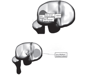
Актуальність. Біомеханічні чинники при ревматоїдному артриті (РА) можуть відігравати важливу роль в ініціюванні та прогресуванні дегенеративних процесів у суглобі, вторинних щодо запального процесу. Проте послідовність біомеханічних і біохімічних процесів, що регулюють ці події in vivo, поки недостатньо ясна. Розуміння величин біомеханічних навантажень на суглобові поверхні в умовах контрактури суглобів нижніх кінцівок при РА і участь у цьому процесі м’язових сил може сприяти розвитку нових поглядів і підходів до лікувальних заходів, специфічних для кожної стадії захворювання. Мета: створити імітаційну комп’ютерну 3D-модель колінного суглоба (КоС) при його згинальній контрактурі на пізніх стадіях РА з метою об’єктивної оцінки функціонального стану суглоба й прилеглих тканин та виявлення перспектив лікування та контролю ефективності реабілітаційних заходів. Матеріали та методи. В основу аналітичних розрахунків покладено дані попередніх досліджень щодо кількісної оцінки контрактури КоС (піддатливість контрактури зовнішній коригуючий дії) та розрахунки суглобових сил, що виникають при ходьбі пацієнта з РА. Визначення напружень та навантажень у КоС проводили при згинально-розгинальній контрактурі 30°. Результати. Для вирішення завдання розроблено декілька розрахункових схем, на яких для більшої наочності зображені всі розміри та діючі сили в натуральну величину для конкретної моделі. Унаслідок примусового пасивного згинання в КоС на 6° навантаження на виростки плато великогомілкової кістки збільшилося на 12,8 %, унаслідок пасивного розгинання в КоС на 3° — на 95,2 %, що є критичним та може призводити до деградації кісткової тканини в ділянці контакту. Висновки. Зростання навантаження на задні відділи плато великогомілкової кістки та, відповідно, напружень у ділянках контакту виростків стегнової кістки з плато великогомілкової кістки може призводити до прогресування клінічної картини РА з посиленням явищ артрофіброзу та остеоартриту саме в задніх відділах КоС.
Background. Rheumatoid arthritis (RA) is an immunomodulatory, chronic inflammatory disease accompanied by the proliferation and articular cartilage destruction that cause disability. Biomechanical factors in RA can play an important role in the onset and progress of the joint degenerative processes, secondary to the inflammation process. The biomechanical factors in RA can play an essential role in the start and progress of the degenerative processes within the joint that are secondary to the inflammatory process. Materials and methods. A solid simulation 3D-model of the knee joint was created that contained both tibia and fibula, the femur bone, femoral condyle cartilage and tibial plateau cartilage, menisci. It was done for further analytical calculations and finite element modeling calculations. Analytical calculations are based on the data of previous studies of quantitative evaluation of the knee joint contracture (compliance of contracture to the external corrective action) and on calculations data of the joint forces that manifest in ambulation of the patient with RA. Results. The created simulation computer 3D-model of a knee joint with its flexion contracture at late stages of RA shows that the forced passive flexion in the knee joint to 6°, the load on the condyles of the tibial plateau increased by 12.8 %, and as a result of forced passive flexion in the knee joint to 3°, the load on the condyles of the tibial plateau increased by 95.2 %, which is critical and may cause degradation of the bone tissue in the contact area. Conclusions. Increase of load on the lateral areas of the tibial plateau and, correspondingly, the tensions on the contact areas of the femur bone condyles with the tibial plateau, may contribute to the progress of the clinical picture of RA with the increase of arthrofibrosis and osteoarthritis events, particularly in the lateral areas of the knee joint.
ревматоїдний артрит; колінний суглоб; згинальна контрактура; математичне моделювання; метод скінченних елементів; напружено-деформуючий стан
rheumatoid arthritis; knee joint; flexion contracture; mathematic modelling; finite element method; load-deformation state
Для ознакомления с полным содержанием статьи необходимо оформить подписку на журнал.
- Watson R.S., Gouze E., Levings P.P., Bush M.L., Kay J.D. et al. Gene delivery of TGF-β1 induces arthrofibrosis and chondrometaplasia of synovium in vivo. Lab. Invest. 2010 Nov. 90(11). 1615-27. doi: 10.1038/labinvest.2010.145.
- Ouyang X., Ghani A., Mehal W.Z. Inflammasome biology in fibrogenesis. Biochim. Biophys. Acta. 2013 Jul. 1832(7). 979-88. doi: 10.1016/j.bbadis.2013.03.020.
- Canovas F., Dagneaux L. Quality of life after total knee arthroplasty. Orthop. Traumatol. Surg. Res. 2018 Feb. 104(1S). S41-S46. doi: 10.1016/j.otsr.2017.04.017.
- Øiestad B.E., Juhl C.B., Eitzen I., Thorlund J.B. Knee extensor muscle weakness is a risk factor for development of knee osteoarthritis. A systematic review and meta-analysis. Osteoarthritis Cartilage. 2015 Feb. 23(2). 171-7. doi: 10.1016/j.joca.2014.10.008.
- Dean C.S., Chahla J., Mikula J.D., Mitchell J.J., LaPrade R.F. Arthroscopic Posteromedial Capsular Release. Arthrosc. Tech. 2016 May 16. 5(3). e495-500. doi: 10.1016/j.eats.2016.01.034.
- Herman M.J., Martinek M.A., Abzug J.M. Complications of tibial eminence and diaphyseal fractures in children: prevention and treatment. J. Am. Acad. Orthop. Surg. 2014 Nov. 22(11). 730-41. doi: 10.5435/JAAOS-22-11-730.
- Dell’Isola A., Smith S.L., Andersen M.S., Steultjens M. Knee internal contact force in a varus malaligned phenotype in knee osteoarthritis (KOA). Osteoarthritis Cartilage. 2017 Dec. 25(12). 2007-2013. doi: 10.1016/j.joca.2017.08.010.
- Werner B.C., Cancienne J.M., Miller M.D., Gwathmey F.W. Incidence of Manipulation Under Anesthesia or Lysis of Adhesions After Arthroscopic Knee Surgery. Am. J. Sports Med. 2015 Jul. 43(7). 1656-61. doi: 10.1177/0363546515578660.
- George J.M. Valgus Deformity Correction in Total Knee Replacement: An Overview. Knee Surgery — Reconstruction and Replacement. IntechOpen. 2020. doi: 10.5772/intechopen.89739.
- Schiavone Panni A., Cerciello S., Vasso M., Tartarone M. Stiffness in total knee arthroplasty. J. Orthop. Traumatol. 2009 Sep. 10(3). 111-8. doi: 10.1007/s10195-009-0054-6.
- Formby P.M., Donohue M.A., Cannova C.J., Caulfield J.P. Hydraulic distension of the knee: a novel treatment for arthrofibrosis after total knee replacement (case series). ANZ J. Surg. 2016 Jun. 86(6). 480-2. doi: 10.1111/ans.13540.
- Khatri K., Bansal D., Rajpal K. Management of Flexion Contracture in Total Knee Arthroplasty [Internet]. Knee Surgery — Reconstruction and Replacement. IntechOpen. 2020. doi: 10.5772/intechopen.90417.
- Kukin I.A., Kirpichjov I.V., Maslov L.B., Vihrev S.V. Particularities of the strength characteristics of spongy bone in diseases of the hip joint. Fundamental Research. 2013. 7. 328-33.
- Ipach I., Mittag F., Lahrmann J., Kunze B., Kluba T. Arthrofibrosis after TKA — influence factors on the absolute flexion and gain in flexion after manipulation under anaesthesia. BMC Musculoskelet. Disord. 2011 Aug 12. 12. 184. doi: 10.1186/1471-2474-12-184.
- Kalson N.S., Borthwick L.A., Mann D.A., Deehan D.J., Lewis P. et al. International consensus on the definition and classification of fibrosis of the knee joint. Bone Joint J. 2016 Nov. 98-B(11). 1479-1488. doi: 10.1302/0301-620X.98B10.37957.
- Pujol N., Boisrenoult P., Beaufils P. Post-trauma–tic knee stiffness: surgical techniques. Orthop. Traumatol. Surg. Res. 2015 Feb. 101(1 Suppl.). S179-86. doi: 10.1016/j.otsr.2014.06.026.
- Бабко А.М., Герасименко А.С., Мазевич В.Б. Механізм формування контрактур кульшового та колінного суглобів на ранніх стадіях ревматоїдного артриту (натурний експеримент). Вісн. ортопедії травматології та протезування. 2019. 4. 57-65. doi:10.37647/0132-2486-2019-103-4-53-61.
- Deshmukh A.J., Rathod P.A., Moses M.J., Snir N., Marwin S.E., Dayan A.J. Does a non-stemmed constrained condylar prosthesis predispose to early failure of primary total knee arthroplasty? Knee Surg. Sports Traumatol. Arthrosc. 2016 Oct. 24(10). 3194-3199. doi: 10.1007/s00167-014-3494-3.