
Журнал «Боль. Суставы. Позвоночник» Том 12, №3, 2022
Вернуться к номеру
Педобарографія — засіб моніторингу процесу функціонального відновлення нестабільних ушкоджень гомілковостопного суглоба
Авторы: Сулима В.С., Філяк Ю.О., Чужак А.В.
Івано-Франківський національний медичний університет, м. Івано-Франківськ, Україна
Рубрики: Ревматология, Травматология и ортопедия
Разделы: Справочник специалиста
Версия для печати
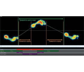
Метод педобарографії дозволяє об’єктивізувати динаміку відновлення функціональної здатності травмованої кінцівки. Метод набуває популярності при визначенні функціональних результатів хірургічного лікування хворих з пошкодженнями гомілковостопного суглоба. Тонкощі педобарографічного моніторингу протягом лікування дають змогу отримати цифрові показники, які при ретельному статистичному аналізі дозволять не тільки суттєво покращити діагностичний процес, але й контролювати процес функціонального одужання хворих з ушкодженнями нижніх кінцівок. Зміни цифрових статичних і динамічних показників педобарографії вказують на прогрес у функціональному відновленні травмованої кінцівки чи необхідність корекції реабілітаційного процесу. Проте існує проблема інтерпретації і конкретизації відомих показників та індексів відповідно до патології. Огляд наукових публікацій, пошук яких виконаний у базах Scopus, Web of Science і The Cochrane Library, покликаний проаналізувати можливості застосування методу педобарографії в діагностиці пошкоджень гомілковостопного суглоба, переломів кісточок гомілки, які за кількісними показниками становлять 20–28 % від усіх переломів кісток людини. Кінематичні особливості організму людини в нормі та при патології слід ретельно аналізувати й практично використовувати в процесі моніторингу відновлення ходи протягом періоду реабілітації в пацієнтів з переломами гомілковостопного суглоба і не тільки. Контроль і виявлення відхилень педобарографічних показників слід детально аналізувати з метою раннього виявлення неусунутої проблеми до появи клінічних ознак хронічної нестабільності. Літературний пошук доводить, що аналіз усього спектра статичних і динамічних показників педобарографії ушкодженої і здорової кінцівок протягом періоду післяопераційного відновлення ходи дає змогу оцінити ефективність реабілітаційних заходів, спрямованих на функціональне відновлення нестабільних пошкоджень гомілковостопного суглоба.
The pedobarography method makes it possible to objectify the dynamics of restoring the functional capacity of an injured limb. The value of the method is gaining popularity in determining the functional results of surgical treatment of the patients with ankle joint injuries. The details of pedobarographic monitoring during the treatment allow to obtain digital indices, which with careful statistical analysis can significantly improve not only the diagnostic process, but also control the process of functional recovery of the patients with injuries of the lower extremities. Changes in quantitative digital static and dynamic indices of pedobarography mean progress in the functional restoration of the injured limb or the need in the correction of the rehabilitation process. However, there is a problem in the interpretation and specification of the known indices and indices in accordance with the pathology. A review of the scientific publications based on the Scopus, Web of Science and The Cochrane Library databases is designed to analyze the possibilities of using the pedobarography method in the diagnosis of ankle joint injuries, tibial bone fractures, which in accordance with quantitative indices make 20-28 % of all fractures of human bones. The kinematic features of the human body in normal and pathological conditions should be carefully analyzed and practically used in the monitoring process of the recovery of walking during the rehabilitation period in the patients with ankle joint fractures and not only. Control and detection of the deviations of pedobarographic indices should be analyzed in details with the aim of early detection of an unresolved problem before the appearance of clinical signs of chronic instability. The literature search proves that the analysis of the entire range of static and dynamic indices of pedobarography of damaged and healthy limbs during the period of postoperative recovery of walking makes it possible to evaluate the effectiveness of rehabilitation measures aimed at functional restoration of unstable injuries of the ankle joint.
огляд; гомілковостопний суглоб; педобарографія; моніторинг; хода
review; ankle joint; pedobarography; monitoring; gait cycle
Для ознакомления с полным содержанием статьи необходимо оформить подписку на журнал.
- Michelson J.D., Magid D., McHale K. Clinicalutility of a stability-base dankle fracture classification system. J. Orthop. Trauma. 2007. 21(5). 307-15. doi: 10.1097/BOT.0b013e318059aea3.
- Dawe E.J., Shafafy R., Quayle J., Gougoulias N., Wee A., Sakellariou A. The effect of different methods of stability assessment onf ixation rate and complication sinsupination external rotation (SER) 2/4 anklefractures. Foot Ankle Surg. 2015. 21(2). 86-90. doi: 10.1016/j.fas.2014.09.010.
- Stufkens S.A., van den Bekerom M.P., Kerkhoffs G.M., Hintermann B., van Dijk C.N. Long-term outcome after 1822 operatively treated anklef ractures: a systematic review of the literature. Injury. 2011. 42(2). 119-27. doi: 10.1016/j.injury.2010.04.006.
- Mehta S.S., Rees K., Cutler L., Mangwani J. Understanding risks and complications in the management of ankle fractures. Indian J. Orthop. 2014 Sep. 48(5). 445-52. doi: 10.4103/0019-5413.139829.
- Shaner A.C., Sirisreetreerux N., Shafiq B., Jones L.C., Hasenboehler E.A. Open versus minimally invasive fixation of a simulated syndesmotic injury in a cadaver model. J. Orthop. Surg. Res. 2017 Oct 27. 12(1). 160. doi: 10.1186/s13018-017-0658-0.
- Bleakley C.M., Matthews M., Smoliga J.M. Mostankle sprain research is either false or clinically unimportant: A 30-year audit of randomized controlled trials. J. Sport Health Sci. 2021 Sep. 10(5). 523-529. doi: 10.1016/j.jshs.2020.11.002.
- Choi Young Rak, Lee Ho Seong, Kim Dong Eun, Lee Dong Ho, Kim Jong Min, Ahn Ji Yong. The Diagnostic Value of Pedobarography. Orthopedics. 2014. 37(12). e1063-67. doi:10.3928/01477447-20141124-52.
- Hagen L., Pape J.P., Kostakev M., Peterlein C.D. Pedobarographic changes during first month after subtalar extra-articular screw arthroereisis (SESA) operation of juvenile flexible flat foot. Arch. Orthop. Trauma Surg. 2020 Mar. 140(3). 313-320. doi: 10.1007/s00402-019-03230-7.
- Цапенко В.В., Терещенко М.Ф., Іваненко Р.О. Біомеханічний метод оцінки ефективності використання індивідуальних ортезів стопи. Вчені записки ТНУ імені В.І. Вернадського. Серія: Технічні науки. 2021. 32(71). 13-19. doi: 10.32838/2663-5941/2021.2-2/03.
- Suciu O., Onofrei R.R., Totorean A.D., Suciu S.C., Amaricai E.C. Gait analysis and functional outcomes after twelve-week rehabilitation in patients with surgically treated ankle fractures. Gait Posture. 2016 Sep. 49. 184-189. doi: 10.1016/j.gaitpost.2016.07.006.
- Fenwick A., Kröger N., Jovic S., Hölscher-Doht S., Meffert R., Jansen H. Pedobarography shows no difference singait after talar fractures. Technol. Health Care. 2020. 28(1). 85-92. doi: 10.3233/THC-191667.
- Genc Y., Gultekin A., Duymus T.M., Mutlu S., Mutlu H., Komur B. Pedobarography in the Assessment of Postoperative Calcaneal Fracture Pressure With Gait. J. Foot Ankle Surg. 2016 Jan-Feb. 55(1). 99-105. doi: 10.1053/j.jfas.2015.07.018.
- Hosoi I., Kobayashi E., Chang S.H. et al. Development of intraoperative plantar pressure measuring system considering weight bearing axis. International Journal of Computer Assisted Radiology and Surgery. 2018. 14(2). 385-395. doi:10.1007/s11548-018-1862-z.
- Horisberger M., Hintermann B., Valderrabano V. Alterations of plantar pressure distribution in post traumaticend-stage ankleosteoarthritis. Clin. Biomech. (Bristol, Avon). 2009 Mar. 24(3). 303-7. doi: 10.1016/j.clinbiomech.2008.12.005.
- Losch A., Meybohm P., Schmalz T. et al. Funktionelle Ergebnisse bei Freizeitsportlern in der dynamischen Ganganalyse 1 Jahr nach operativ versorgten Sprunggelenkfrakturen. Sportverletz Sportschaden 2002. 16(3). 101-107. doi: 10.1055/s-2002-34750.
- de Kruijff L.G.M., Prins M., van der Krans A., Hoencamp R., van der Wurff P. Combat-related foot injuries: impact on gait and functional outcome. J. R. Army Med. Corps. 2018 Sep. 164(5). 322-327. doi: 10.1136/jramc-2017-000870.
- Schmiegel A., Rosenbaum D., Schorat A., Hilker A., Gaubitz M. Assessment of foot impairment in rheumatoid arthritis patients by dynamic pedobarography. Gait Posture. 2008 Jan. 27(1). 110-4. doi: 10.1016/j.gaitpost.2007.02.008.
- Lorkowski J., Gawronska K., Pokorski M. Pedobarography: A Review on Methods and Practical Use in Foot Disorders. Appl. Sci. 2021. 11. 11020. doi: 10.3390/app112211020.
- Lee S.H., Lee O.S., Teo S.H., Lee Y.S. Change in gait after high tibial osteotomy: A systematic review and meta-analysis. Gait Posture. 2017 Sep. 57. 57-68. doi: 10.1016/j.gaitpost.2017.05.023.
- Kim S.G., Nam C.W., Yong M.S. The effect of increase in baggage weight on elderly women's lower extremity muscle activation during gait. Arch. Gerontol. Geriatr. 2014 Nov-Dec. 59(3). 574-6. doi: 10.1016/j.archger.2014.07.015.
- Bowen T.R., Miller F., Castagno P., Richards J., Lipton G. A method of dynamic foot-pressure measurement for the evaluation of pediatric orthopaedic foot deformities. J. Pediatr. Orthop. 1998 Nov-Dec. 18(6). 789-93. PMID: 9821137.
- Schmiegel A., Rosenbaum D., Schorat A., Hilker A., Gaubitz M. Assessment of foot impairment in rheumatoid arthritis patients by dynamic pedobarography. Gait Posture. 2008 Jan. 27(1). 110-4. doi: 10.1016/j.gaitpost.2007.02.008.
- Frigg A., Frigg R., Wiewiorski M., Goldoni J., Horisberger M. Facilitating the interpretation of pedobarography: the relative midfoot index as marker for pathologic gait in ankle osteoarthritic and contralateral feet. J. Foot Ankle Res. 2016. 9(47). n.a. doi: 0.1186/s13047-016-0177-y.
- Duval K., Lam T., Sanderson D. The mechanical relationship between the rearfoot, pelvis and low-back. Gait Posture. 2010 Oct. 32(4). 637-40. doi: 10.1016/j.gaitpost.2010.09.007.
- Buldt A.K., Forghany S., Landorf K.B., Levinger P., Murley G.S., Menz H.B. Foot posture is associated with plantar pressure during gait: A comparison of normal, planus and cavus feet. Gait Posture. 2018 May. 62. 235-240. doi: 10.1016/j.gaitpost.2018.03.005.
- Woźniacka R., Oleksy Ł., Jankowicz-Szymańska A., Mika A., Kielnar R., Stolarczyk A. The association between high-arched feet, plantar pressure distribution and body posture in young women. Sci Rep. 2019 Nov 20. 9(1). 17187. doi: 10.1038/s41598-019-53459-w.
- Crenshaw S.J., Pollo F.E., Brodsky J.W. The effect of ankle position on plantar pressure in a short leg walking boot. Foot Ankle Int. 2004 Feb. 25(2). 69-72. doi: 10.1177/107110070402500206.
- Monteiro R.L., Sartor C.D., Ferreira J.S.S.P., Dantas M.G.B., Bus S.A., Sacco I.C.N. Protocol for evaluating the effects of a foot-ankle therapeutic exercise program on daily activity, foot-ankle functionality, and biomechanics in people with diabetic polyneuropathy: a randomized controlled trial. BMC Musculoskelet. Disord. 2018 Nov 14. 19(1). 400. doi: 10.1186/s12891-018-2323-0.
- Aiona M., Do K.P., Emara K., Dorociak R., Pierce R. Gait patterns in children with limb length discrepancy. J. Pediatr. Orthop. 2015 Apr-May. 35(3). 280-4. doi: 10.1097/BPO.0000000000000262.
- Tenenbaum S., Coleman S.C., Brodsky J.W. Improvement in gait following combined ankle and subtalar arthrodesis. J. Bone Joint Surg. Am. 2014 Nov 19. 96(22). 1863-9. doi: 10.2106/JBJS.M.01448.
- Manjra M.A., Naude J., Birkholtz F., Glatt V., Tetsworth K., Hohmann E. The relationship between gait and functional outcomes in patients treated with circular external fixation for malunited tibial fractures. Gait Posture. 2019 Feb. 68. 569-574. doi: 10.1016/j.gaitpost.2019.01.008.
- McNab B., Sadler S., Lanting S., Chuter V. The relationship between foot and ankle joint flexibility measures and barefoot plantar pressures in healthy older adults: a cross-sectional study. BMC Musculoskelet. Disord. 2022 Jul 30. 23(1). 729. doi: 10.1186/s12891-022-05618-w.
- Buldt A.K., Allan J.J., Landorf K.B., Menz H.B. The relationship between foot posture and plantar pressure during walking in adults: A systematic review. Gait Posture. 2018 May. 62. 56-67. doi: 10.1016/j.gaitpost.2018.02.026.
- Mirando M., Conti C., Zeni F., Pedicini F., Nardone A., Pavese C. Gait Alterations in Adults after Ankle Fracture: A Systematic Review. Diagnostics (Basel). 2022 Jan 14. 12(1). 199. doi: 10.3390/diagnostics12010199.
- Hopkins J.T., Coglianese M., Glasgow P., Reese S., Seeley M.K. Alterations in evertor/invertor muscle activation and center of pressure trajectory in participants with functional ankle instability. J. Electromyogr. Kinesiol. 2012 Apr. 22(2). 280-5. doi: 10.1016/j.jelekin.2011.11.012.
- Zhang X., Li B. Influence of in-shoe heel lifts on plantar pressure and center of pressure in the medial-lateral direction during walking. Gait Posture. 2014 Apr. 39(4). 1012-6. doi: 10.1016/j.gaitpost.2013.12.025.
- Francis C.A., Lenz A.L., Lenhart R.L., Thelen D.G. The modulation of forward propulsion, vertical support, and center of pressure by the plantarflexors during human walking. Gait Posture. 2013 Sep. 38(4). 993-7. doi: 10.1016/j.gaitpost.2013.05.009.
- Chevalier T.L., Hodgins H., Chockalingam N. Plantar pressure measurements using an in-shoe system and a pressure platform: a comparison. Gait Posture. 2010 Mar. 31(3). 397-9. doi: 10.1016/j.gaitpost.2009.11.016.
- Jeon E.T., Cho H.Y. A Novel Method for Gait Analysis on Center of Pressure Excursion Based on a Pressure-Sensitive Mat. Int. J. Environ. Res. Public Health. 2020 Oct 26. 17(21). 7845. doi: 10.3390/ijerph17217845.
- van Mierlo M., Vlutters M., van Asseldonk E.H.F., van der Kooij H. Centre of pressure modulations in double support effectively counteract anteroposterior perturbations during gait. J. Biomech. 2021 Sep 20. 126. 110637. doi: 10.1016/j.jbiomech.2021.110637.
- Bamber Z.A., Wheeler P.C., He X., Ling S.K.K., Yung P.S.H., Fong D.T.P. Screening for laterally deviated plantar pressure during stance using the Cumberland ankle instability tool and anthropometric measures. Res. Sports Med. 2021 Jul-Aug. 29(4). 323-335. doi: 10.1080/15438627.2020.1857250.
- Honert E.C., Hoitz F., Blades S., Nigg S.R., Nigg B.M. Estimating Running Ground Reaction Forces from Plantar Pressure during Graded Running. Sensors (Basel). 2022 Apr 27. 22(9). 3338. doi: 10.3390/s22093338.
- Arno F., Roman F., Martin W., Jennifer G., Monika H. Facilitating the interpretation of pedobarography: the relative midfoot index as marker for pathologic gait in ankle osteoarthritic and contralateral feet. J. Foot Ankle Res. 2016 Dec 1. 9. 47. doi: 10.1186/s13047-016-0177-y.
- Espy D.D., Yang F., Bhatt T., Pai Y.C. Independent influence of gait speed and step length on stability and fall risk. Gait Posture. 2010 Jul. 32(3). 378-82. doi: 10.1016/j.gaitpost.2010.06.013.
- Voss S., Joyce J., Biskis A. et al. Normative database of spatiotemporal gait parameters using inertial sensors in typically developing children and young adults. Gait Posture. 2020 Jul. 80. 206-213. doi: 10.1016/j.gaitpost.2020.05.010.
- Hayashi K., Fukuyasu-Matsuo S., Inoue T. et al. Effects of cyclic stretching exercise on long-lasting hyperalgesia, joint contracture, and muscle injury following cast immobilization in rats. Physiol. Res. 2020. 69(5). 861-70. doi: 10.33549/physiolres.934437.
- Kahn J.H., Hornby T.G. Rapid and long-term adaptations in gait symmetry following unilateral step training in people with hemiparesis. Phys. Ther. 2009 May. 89(5). 474-83. doi: 10.2522/ptj.20080237.
- Reisman D.S., McLean H., Keller J., Danks K.A., Bastian A.J. Repeated split-belt treadmill training improves poststroke step length asymmetry. Neurorehabil. Neural. Repair. 2013 Jun. 27(5). 460-8. doi: 10.1177/1545968312474118.
- Yen S.C., Schmit B.D., Wu M. Using swing resistance and assistance to improve gait symmetry in individuals post-stroke. Hum. Mov. Sci. 2015 Aug. 42. 212-24. doi: 10.1016/j.humov.2015.05.010.
- Sheikh M., Azarpazhooh M.R., Hosseini H.A. Randomized comparison trial of gait training with and without compelled weight-shift therapy in individuals with chronic stroke. Clin. Rehabil. 2016 Nov. 30(11). 1088-1096. doi: 10.1177/0269215515611467.
- Lewek M.D., Braun C.H., Wutzke C., Giuliani C. The role of movement errors in modifying spatiotemporal gait asymmetry post stroke: a randomized controlled trial. Clin. Rehabil. 2018 Feb. 32(2). 161-172. doi: 10.1177/0269215517723056.
- Lewek M.D., Bradley C.E., Wutzke C.J., Zinder S.M. The relationship between spatiotemporal gait asymmetry and balance in individuals with chronic stroke. J. Appl. Biomech. 2014 Feb. 30(1). 31-6. doi: 10.1123/jab.2012-0208.
- Judge J.O., Davis R.B. 3rd, Ounpuu S. Step length reductions in advanced age: the role of ankle and hip kine–tics. J. Gerontol. A. Biol. Sci Med. Sci. 1996 Nov. 51(6). M303-12. doi: 10.1093/gerona/51a.6.m303.
- Mehdikhani M., Taylor S., Shideler B.L., Ogrin R., Begg R. Age effects on step adaptation during treadmill walking with continuous step length biofeedback. Gait Posture. 2020 Jul. 80. 174-177. doi: 10.1016/j.gaitpost.2020.04.027.
- Ræder B.W., Stake I.K., Madsen J.E. et al. Randomized trial comparing suture button with single 3.5 mm syndesmotic screw for ankle syndesmosis injury: similar results at 2 years. Acta Orthop. 2020. 91(6). 770-5. doi: 10.1080/17453674.2020.1818175.
- Padmanabhan P., Rao K.S., Gulhar S., Cherry-Allen K.M., Leech K.A., Roemmich R.T. Persons post-stroke improve step length symmetry by walking asymmetrically. J. Neuroeng. Rehabil. 2020 Aug 3. 17(1). 105. doi: 10.1186/s12984-020-00732-z.
- Gama G.L., Savin D.N., Keenan T., Waller S.M., Whitall J. Comparing the effects of adapting to a weight on one leg during treadmill and overground walking: A pilot study. Gait Posture. 2018 Jan. 59. 35-39. doi: 10.1016/j.gaitpost.2017.09.025.
- Kim S.G., Hwangbo G. The effect of obstacle gait training on the plantar pressure and contact time of elderly women. Arch. Gerontol. Geriatr. 2015 May-Jun. 60(3). 401-4. doi: 10.1016/j.archger.2015.02.007.
- Lauzière S., Miéville C., Betschart M., Duclos C., Aissaoui R., Nadeau S. A more symmetrical gait after split-belt treadmill walking increases the effort in paretic plantar flexors in people post-stroke. J. Rehabil. Med. 2016. 48. 576-82. doi: 10.2340/16501977-2117.
- Koldenhoven R.M., Jaffri A.H., DeJong A.F. et al. Gait biofeedback and impairment-based rehabilitation for chronic ankle instability. Scand. J. Med. Sci Sports. 2021 Jan. 31(1). 193-204. doi: 10.1111/sms.13823.
- Eliks M., Ostiak-Tomaszewska W., Lisiński P., Koczewski P. Does structural leg-length discrepancy affect postural control? Preliminary study. BMC Musculoskelet. Disord. 2017. 18(1). 346. doi: org/10.1186/s12891-017-1707-x.
- Sung P.S. Increased double limb support times during walking in right limb dominant healthy older adults with low bone density. Gait Posture. 2018 Jun. 63. 145-149. doi: 10.1016/j.gaitpost.2018.04.036.
- Bautmans I., Jansen B., Van Keymolen B., Mets T. Reliability and clinical correlates of 3D-accelerometry based gait analysis outcomes according to age and fall-risk. Gait Posture. 2011 Mar. 33(3). 366-72. doi: 10.1016/j.gaitpost.2010.12.003.
- Cibulka M.T., Winters K., Kampwerth T. et al. Predicting Foot Progression Angle During Gait Using Two Clinical Measures In Healthy Adults, A Preliminary Study. Int. J. Sports Phys. Ther. 2016 Jun. 11(3). 400-8. PMID: 27274426; PMCID: PMC4886808.
- Elbaz A., Mor A., Segal O. et al. Can single limb support objectively assess the functional severity of knee osteoarthritis? Knee. 2012 Jan. 19(1). 32-5. doi: 10.1016/j.knee.2010.12.004.
- Skvortsov D., Kaurkin S., Prizov A., Altukhova A., Troitskiy A., Lazko F. Biomechanical Changes in Gait Patterns of Patients with Grade II Medial Gonarthritis. Diagnostics (Basel). 2021 Jul 12. 11(7). 1242. doi: 10.3390/diagnostics11071242.
- Schafer Z.A., Perry J.L., Vanicek N. A personalised exercise programme for individuals with lower limb amputation reduces falls and improves gait biomechanics: A block randomised controlled trial. Gait Posture. 2018 Jun. 63. 282-289. doi: 10.1016/j.gaitpost.2018.04.030.
- Remelius J.G., van Emmerik R.E. Time-To-Contact Analysis of Gait Stability in the Swing Phase of Walking in People With Multiple Sclerosis. Motor Control. 2015 Oct. 19(4). 289-311. doi: 10.1123/mc.2013-0106.
- Studenski S., Perera S., Patel K. et al. Gait speed and survival in older adults. JAMA. 2011 Jan 5. 305(1). 50-8. doi: 10.1001/jama.2010.1923.