Інформація призначена тільки для фахівців сфери охорони здоров'я, осіб,
які мають вищу або середню спеціальну медичну освіту.

Підтвердіть, що Ви є фахівцем у сфері охорони здоров'я.


Коморбідний ендокринологічний пацієнт

Коморбідний ендокринологічний пацієнт

Международный эндокринологический журнал Том 18, №4, 2022

Вернуться к номеру

Йодний дефіцит і поширеність вузлового зоба в Україні

Авторы: Tovkai A.O.
Bogomolets National Medical University, Kyiv, Ukraine

Рубрики: Эндокринология

Разделы: Справочник специалиста

Версия для печати


Резюме

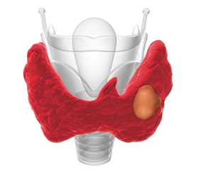
Вузловий зоб діагностується в однієї десятої частини населення світу. Значні відмінності в поширеності захворювань щитоподібної залози між популяціями зумовлені генетичними й екологічними чинниками. Серед останніх найважливішим фактором ризику є дефіцит йоду. Вузловий зоб переважно відзначається в регіонах з йодним дефіцитом. У країнах, які страждають від дефіциту йоду, повідомляється про більші обсяги щитоподібної залози й вищу частоту вузлового зоба. Йодний дефіцит — проблема, актуальна на всій території України. В умовах відсутності масової йодної профілактики в населення відзначається висока частота йододефіцитних захворювань (ЙДЗ). Поширеність зоба в дітей загалом в Україні, навіть за даними офіційної статистики, перевищує 5% бар’єр. Фактична поширеність тиреоїдної патології значно вища за дані у звітах медичних установ, а рівень медіани йодурії відповідає в деяких регіонах йодному дефіциту легкого ступеня. Вирішити проблему профілактики ЙДЗ у населення можна прийняттям на законодавчому рівні постанови про обов’язкове йодування харчової солі в країні, а також проведенням індивідуальної профілактики препаратами калію йодиду в групах особливого ризику. В огляді проведена оцінка поширеності вузлового зоба в Україні порівняно з іншими країнами на тлі йодного дефіциту. У 2016 році в Україні поширеність вузлового зоба становила 707,8 на 100 тис. населення, потім через 5 років цей показник зріс до 891,5 випадку (+25,9 %). Показники захворюваності на вузловий зоб у країні у 2016 році становили 71,9, а на кінець 2020 року — 90,2 на 100 тис. населення (+25,5 %). На основі результатів Фремінгемського дослідження оцінений ризик розвитку вузлового зоба протягом життя, він становить 5–10 %.

More than one tenth of the world population is to some degree affected by goitre and most of these harbour nodules. The large differences in thyroid disease prevalence between populations may be caused by genetic and environmental factors. Among the latter, iodine deficiency seems by far to be the most important risk factor. Thus, nodular goitre is a condition predominantly seen in iodine deficient areas of the world. Large thyroid volumes and high frequencies of goitres have been reported in countries affected by iodine deficiency. In the present review, we evaluated prevalences of thyroid nodules in iodine-deficient countries. In 2016 in Ukraine the prevalence of nodular goiter was 707.8 per 100 thousand population, then after 5 years the figure slowly increased to 891.5 cases, respectively (+25.9 %). The existing high indices of newly diagnosed patients with nodular goitre in the country in 2016 were 71.9, and at the end of 2020 — 90.2 initially established cases per 100 thousand population, with a positive increase of +25.5 %. In the Whickham survey, 20 % of women and 5 % of men who had goitres in the initial survey showed no evidence of goitre in a follow-up survey. An average growth rate in the multinodular goitre of 5–20 % was reported in iodine-sufficient areas. On the basis of the results of the Framingham survey, the estimated lifetime risk for developing a nodule is 5–10 %. Thyroid nodule size can increase, decrease, or remain stable, and thyroid nodules may eventually also disappear over time. Solid nodules more frequently increase, whereas cystic nodules can shrink or disappear. If the goitre has been present for some time, autonomous function of the nodules and eventually hyperthyroidism develop. The rate of progression from euthyroidism to subclinical and overt hyperthyroidism is about 10 %.


Ключевые слова

йодний дефіцит; вузловий зоб; епідеміологія; огляд

iodine deficiency; nodular goitre; epidemiology; review

One of the priority tendencies of health care in most countries is the prevention of iodine deficiency disorders (IDD), i.e. all pathological conditions that develop in the population as a result of iodine deficiency (ID), which can be prevented with normal iodine intake. Thyroid pathology is one of the most common reasons for the population to seek medical attention, due to its high prevalence in iodine-deficient and contaminated areas, as well as the availability of the research methods [1]. Iodine is necessary for the production of adequate amounts of the thyroid hormones. The daily requirement of iodine depends on age and physiological condition and ranges from 90 to 250 μg/day [1, 2].
The spectrum of thyroid diseases is wide, ranging from minor structural changes not influencing the life of the patients to a variety of disorders that may reduce the quality of life and in some circumstances also affect life expectancy [3]. Among the mild but frequent disorders are goitre and benign thyroid nodules. Nodules, goitre, and nodular goitre still remain common thyroid disorders. These conditions often develop simultaneously, but many subjects harbour nodules within the thyroid gland without development of goitre, and in populations with excessive iodine intake goitres are often without nodules [4].
Various definitions of goitre have been used during the last hundred years depending on which study methods have been available. After the introduction of thyroid ultrasound examination, the definition of goitre has been based on the measurement of the total thyroid volume using various principles. Goitre is now defined as a total thyroid volume exceeding the mean size +3 standard deviations in iodine-replete adult populations, which corresponds to a total volume larger than 18 cm3 in women and 25 cm3 in men [5]. The prevalence of goitre observed in a study may depend not only on the thyroid size, but also on the upper reference limit definition. Evidently, it may be difficult to compare goitre prevalences between studies. 
Nodular goitre is a recognizable thyroid enlargement characterised by excessive growth and structural and/or functional transformation of one or several areas within the normal thyroid tissue [6].
We explore the role of iodine intake level as a risk factor for goitre and nodules. The prevalence of goitre and intrathyroidal nodules may depend on several factors [7]. Among the environmental factors investigated, low iodine intake is a major cause for nodular goitre [8]. 
During 1990 to 1999 years, the overall goitre prevalence was estimated to be unchanged from 12 to 13 %, but the criteria for defining goitre had changed from palpation towards the more precise ultrasonographical criteria [9]. 
Iodine intake is a major determinant for thyroid volumes. The association between thyroid size and iodine intake is furthermore supported by studies comparing areas with different iodine intake using same study design. 
The prevalence of nodular goitre in various regions of the World differing in iodine status also associates negatively with urinary iodine excretion.
According to the WHO, about 2 billion people live under conditions of chronic ID [1]. Iiodine deficiency is one of the most common causes of mental retardation. In recent years, studies all round the world have shown that the average level of intellectual development (IQ) in areas with severe iodine deficiency is 15–20 % lower than in areas with iodine deficiency [10, 11]. 
Radioactive iodine, flying out of the destroyed reactor of the Chernobyl nuclear power plant, like a corona virus quickly spread throughout Europe. Iodine deficiency in the regions of Ukraine adjacent to nuclear power plants became a factor in increased uptake of radioactive iodine by the thyroid gland in children and after 5 years led to an epidemic of thyroid cancer [12]. Optimal iodine intake could become a kind of vaccination, which sharply reduces the risk of developing thyroid cancer, as has happened after the accident at the Fukushima nuclear power plant in Japan. 
Endemic goiter was eliminated 40 years ago, but returned to the country in the early 1990s after the collapse of iodized salt production and has not been eliminated to this day due to the lack of a legislative framework for mandatory salt iodization. The actual average consumption of iodine by residents of Ukraine is from 40 to 80 mcg per day mild and moderate iodine deficiency was detected throughout Ukraine [13]. 
According to WHO, over the past 20 years, iodine deficiency has been eliminated in 115 countries of the world, and the number of iodine-deficient countries has dropped to 25, but Ukraine is still among them. 
The medical and social significance and urgency of the problem of IDD in Ukraine is caused by the fact that in almost all regions there is a natural deficiency of iodine. Traditionally in Ukraine, the regions with severe iodine deficiency include Zakarpattia, Ivano-Frankivsk, Volyn, Lviv, Rivne, Chernivtsi and Ternopil regions. The absolute number of cases of thyroid diseases in Ukraine over the past 10 years has increased from 689 thousand to 1 million 846 thousand, in terms of prevalence, respectively, 941.6 and 4210.4 cases per 100 thousand population. 
Nodular goitre is one of the forms of thyroid pathology, which causes constant clinical interest due to the threat of malignancy of these tumours. The dynamics of epidemiological observations on the prevalence of nodular goitre in Ukraine is characterized by an increase in the prevalence and incidence of nodular goitre over the past five years [14]. In 2016 in Ukraine the prevalence of nodular goiter was 707.8 per 100 thousand population, then after 5 years the figure slowly increased to 891.5 cases, respectively (+25.9 %) [15]. The existing high indices of newly diagnosed patients with nodular goitre in the country in 2016 were 71.9, and at the end of 2020 — 90.2 initially established cases per 100 thousand population, with a positive increase of +25.5 % in terms of clarity [15]. 
In comparative study [16], 40 adults without goiter (control group) and 16 adults with diagnosed nodular goiter (NG) were examined. Inductively coupled plasma optical emission spectrometry (ICP-OES) was used for the measurements of Mg, Ca, Se, Zn, Cu, and Fe in serum of patients with NG and control group. Patients with nodular goiter had lower serum values of Ca, Mg, Se, Cu, Fe, and Zn than those in the control group. The presence of mild iodine deficiency was evident in both groups with the median urinary iodine excretion (UIE) 80.5 μg/L in the control group and 64.5 μg/L in goiter group. There was a positive association between goiter presence and low concentration of Ca in serum (odds ratio (OR) = 2.29 (1.26–3.55), p < 0.05) in the NG group. High relative risk of goiter was observed at low concentrations of magnesium (OR = 3.33 (1.39–7.62), p < 0.05) and selenium (OR = 1.63, (1.16–1.78), p < 0.05) in comparison with OR values in the control group. Low concentrations of Ca, Mg, Zn, and Se in serum combined with reduced UIE resulted in the highest risk of goiter (OR = 12.5, (2.15–79.42), p < 0.01). This study proved that thyroglobulin concentration in serum is the reliable indicator of nodular goiter. We also suggest that a combination of low concentrations of Ca, Mg, Zn, Cu, and Se in blood serum, and reduced iodine concentration in urine resulted in the highest risk of nodular goiter development [16].
The large differences in thyroid disease prevalence between populations may be caused by genetic and environmental factors. Among the latter, iodine deficiency seems by far to be the most important risk factor. Thus, nodular goitre is a condition predominantly seen in iodine deficient areas of the world. 
A few epidemiological studies have evaluated the clinical evolution of goitre. In the Whickham survey [17], 20 % of women and 5 % of men who had goitres in the initial survey showed no evidence of goitre in a follow-up survey [17]. An average growth rate in the multinodular goitre of 5–20 % was reported in iodine-sufficient areas [18]. On the basis of the results of the Framingham survey, the estimated lifetime risk for developing a nodule is 5–10 % [19]. Thyroid nodule size can increase, decrease, or remain stable, and thyroid nodules may eventually also disappear over time. Solid nodules more frequently increase, whereas cystic nodules can shrink or disappear. If the goitre has been present for some time, autonomous function of the nodules and eventually hyperthyroidism develop [20]. The rate of progression from euthyroidism to subclinical and overt hyperthyroidism is about 10 %. 
In a Dutch study of 90 people with euthyroid multinodular goitre, mainly women with a mean age of 55 years, eight became hyperthyroid within 7 years, and all of them had autonomous function before becoming hyperthyroid [21]. In a study conducted at the Mayo Clinic, 60 % of people with multi-nodular goitre over 60 years had thyrotoxicosis [22]. The average duration of the goitre before the onset of thyrotoxicosis was 17 years; the longer the goitre had been present, the greater the tendency for thyrotoxicosis to develop. Iodine intake has been shown to modulate the pattern of thyroid diseases in cross-sectional studies comparing populations living in areas characterized by different iodine intake. 
In iodine-deficient countries, non-autoimmune hyperthyroidism is more frequent, being the natural evolution of goitre, the development of thyroid autonomy, and eventually of thyrotoxicosis. On the other hand, studies in populations living in areas with varying iodine intakes, in the UK, Denmark, and Iceland, have shown that the frequency of thyroid autoimmunity and of hypothyroidism is higher in iodine replete than in iodine deficient populations [23].
In the DanThyr study comparing mild and moderate iodine deficiency, it was found higher thyroid volumes in the area most iodine deficient, whereas no differences in nodule prevalences were reported between the two areas under study [24]. In this study, methods were carefully adjusted to uniformity and sub-populations only differed slightly in degree of iodine deficiency. A span between 10 % prevalence of thyroid nodules in a German study by C. Struve and J. Hinrichs [25] to 32 % in a Danish study performed by N. Knudsen et al. [9] has been observed. Several reasons can be proposed for such a large discrepancy between studies investigating populations with iodine insufficiency. It must be emphasized that ecological conclusions may be hampered by interobserver variation [26], varying population ages and gender compositions of the study cohorts, subjectivity to ultrasonography, and the skills of the ultrasonographers. Best interobserver agreement is on thyroid volume, somewhat lower on nodules [27]. A high interobserver variation of up to 55 % may lead to disagreement on the presence on thyroid nodules [28], and the variation is even larger on echogenicity. Ultrasound is a better tool for investigation of thyroid size than of intrathyroidal structures. Ultrasound may not detect all nodules as shown by D.D. Stark et al. [29] who reported that up to 12 % of intrathyroidal lesions were missed by thyroid ultrasound.
Insulin resistance has been associated with increased thyroid volume and thyroid nodularity. Independently from obesity, insulin resistance is a strong determinant of thyroid volume and thyroid nodularity, with nodularity in 53 % of participants with insulin resistance and 19 % without insulin resistance [30]. This was confirmed with similar results for individuals with metabolic syndrome in an iodine-deficient area [31]. Subsequently, the same research group has, in a prospective setting, investigated thyroid volumes and nodularity in people with diabetes mellitus (DM) or pre-diabetes compared with controls, and found a considerable elevation of nodule prevalence and thyroid volume in DM patients compared with controls (62 % nodules vs 24 % and 20 cm3 vs 11.4 cm3), persons with pre-diabetes having values in-between but closest to type 2 DM [32].
Two studies have investigated the effect of metformin on goitre and nodule prevalence in people with insulin resistance. Insulin resistance was reduced with metformin, and a significant reduction in thyroid nodule volumes was found with metformin but not with levothyroxine treatment [33]. This was confirmed in a study with type 2 DM where metformin treatment was associated with lower occurrence of goitre compared with patients receiving other treatments both in a cross-sectional and in a longitudinal analysis [34].
A number of different explanations for this association have been proposed. Insulin is a potent growth stimulator of thyroid cells in vitro. In contrast, the increased thyroid volume and nodularity could be secondary to an increase in serum thyroid stimulating hormone, with insulin resistance mediated through leptin or numerous other possible pathways, although differences in thyroid stimulating hormone are not a constant finding. Further, a common genetic disposition for insulin resistance and nodular goitre is possible; polymorphisms, including the GLUT genes, have been suggested [35, 36].
In summary, correction of iodine deficiency in adult populations, irrespective of severity, reduces mean thyroid size and the prevalence of diffuse goitre at all ages within a few years. Although iodine repletion usually does not reduce the prevalence of thyroid nodularity in adults older than about 50 years because of largely irreversible fibrotic changes in nodules, it does reduce the risk for development of nodular disease in younger adults.
 
Received 03.05.2022
Revised 14.05.2022
Accepted 17.05.2022

Список литературы

  1. Zimmermann M.B., Boelaert K. Iodine deficiency and thyroid disorders. Lancet Diabetes Endocrinol. 2015. 3(4). 286-95. doi: 10.1016/S2213-8587(14)70225-6. 
  2. Gunnarsdottir I., Dahl L. Iodine intake in human nutrition: a systematic literature review. Food Nutr. Res. 2012. 56. doi: 10.3402/fnr.v56i0.19731. 
  3. Kelley S. Endocrinology Update: Thyroid Disorders. FP Essent. 2016. 451. 11-16. PMID: 27936530.
  4. Jiang H., Tian Y., Yan W., Kong Y., Wang H., Wang A., Dou J. et al. The Prevalence of Thyroid Nodules and an Analysis of Related Lifestyle Factors in Beijing Communities. Int. J. Environ. Res. Public Health. 2016. 13(4). 442. doi: 10.3390/ijerph13040442. 
  5. Miccoli P., Minuto M.N., Orlandini C., Galleri D., Massi M., Berti P. Ultrasonography estimated thyroid volume: a prospective study about its reliability. Thyroid. 2006. 16(1). 37-9. doi: 10.1089/thy.2006.16.37. 
  6. Tonacchera M., Pinchera A., Vitti P. Assessment of nodular goitre. Best Pract. Res. Clin. Endocrinol. Metab. 2010. 24(1). 51-61. doi: 10.1016/j.beem.2009.08.008. 
  7. Venkatesh N., Ho J.T. Investigating thyroid nodules. Aust. Prescr. 2021. 44(6). 200-204. doi: 10.18773/austprescr.2021.055. 
  8. Lou X., Wang X., Wang Z., Mao G., Zhu W., Wang Y. et al. The Effect of Iodine Status on the Risk of Thyroid Nodules: A Cross-Sectional Study in Zhejiang, China. Int. J. Endocrinol. 2020 Aug 18. 2020. 3760375. doi: 10.1155/2020/3760375. 
  9. Knudsen N., Bülow I., Jorgensen T., Laurberg P., Ovesen L., Perrild H. Goitre prevalence and thyroid abnormalities at ultrasonography: a comparative epidemiological study in two regions with slightly different iodine status. Clin. Endocrinol. (Oxf.). 2000. 53(4). 479-85. doi: 10.1046/j.1365-2265.2000.01121.x. 
  10. Niwattisaiwong S., Burman K.D., Li-Ng M. Iodine deficiency: Clinical implications. Cleve Clin. J. Med. 2017. 84(3). 236-244. doi: 10.3949/ccjm.84a.15053. 
  11. Levie D., Korevaar T.I.M., Bath S.C., Murcia M., Dineva M., Llop S., Espada M. et al. Association of Maternal Iodine Status With Child IQ: A Meta-Analysis of Individual Participant Data. J. Clin. Endocrinol. Metab. 2019. 104(12). 5957-5967. doi: 10.1210/jc.2018-02559. 
  12. Tronko M., Mabuchi K., Bogdanova T., Hatch M., Likhtarev I., Bouville A. et al. Thyroid cancer in Ukraine after the Chernobyl accident (in the framework of the Ukraine-US Thyroid Project). J. Radiol. Prot. 2012. 32(1). N65-9. doi: 10.1088/0952-4746/32/1/N65. 
  13. Кravchenko V. Chornobyl Accident and Iodine Deficiency as Risk Factors of Thyroid Pathology in Population of the Affected Regions of Ukraine. International Journal of Endocrinology (Ukraine). 2016. 2(74). 13-20. https://doi.org/10.22141/2224-0721.2.74.2016.70911.
  14. Kamyshna I.I., Pavlovich L.B., Maslyanko V.A., Chornenka Zh.A. Epidemiological assessment of dymamics of the prevalence and incidence of the thyroid gland diseases in Ukraine and Chernivtsi region. Сlinical and Experimental Pathology. 2021. 20(3). 75-81. DOI: 10.24061/1727-4338. XX.3.77.2021.11
  15. Ткаченко В.І., Максимець Я.А., Видиборець Н.В., Коваленко О.Ф. Аналіз поширеності тиреоїдної патології і смертності від неї серед населення Київської області та України за 2007–2017 рр. Міжнародний ендокринологічний журнал. 2018. 14(3). 272-7. doi: https://doi.org/10.22141/2224-0721.14.3.2018.136426.
  16. Kravchenko V.I., Andrusyshyna I.M., Luzanchuk I.A., Polumbryk M.O., Tarashchenko Y.M. Association Between Thyroid Hormone Status and Trace Elements in Serum of Patients with Nodular Goiter. Biol. Trace Elem. Res. 2020. 196(2). 393-399. doi: 10.1007/s12011-019-01943-9. 
  17. Vanderpump M.P., Tunbridge W.M., French J.M. et al. The incidence of thyroid disorders in the community: a twenty-year follow-up of the Whickham Survey. Clin. Endocrinol. (Oxf.). 1995. 43. 55-68. https://doi.org/10.1111/j.1365-2265.1995.tb01894.x.
  18. Rallison M.L., Dobyns B.M., Meikle A.W., Bishop M., Lyon J.L., Stevens W. Natural history of thyroid abnormalities: Prevalence, incidence, and regression of thyroid diseases in adolescents and young adults. The American Journal of Medicine. 1991. 91(4). 363-370. https://doi.org/10.1016/0002-9343(91)90153-O.
  19. Ghassi D., Donato A. Evaluation of the thyroid nodule. Postgrad. Med. J. 2009. 85(1002). 190-5. doi: 10.1136/pgmj.2008.072140. 
  20. Durante C., Grani G., Lamartina L., Filetti S., Mandel S.J., Cooper D.S. The Diagnosis and Management of Thyroid Nodules: A Review. JAMA. 2018. 319(9). 914-924. doi: 10.1001/jama.2018.0898. 
  21. Hooft L., Hoekstra O.S., Boers M., Van Tulder M.W., Van Diest P., Lips P. Practice, efficacy, and costs of thyroid nodule evaluation: a retrospective study in a Dutch university hospital. Thyroid. 2004. 14(4). 287-93. doi: 10.1089/105072504323030942. 
  22. Tamhane S., Gharib H. Thyroid nodule update on diagnosis and management. Clin. Diabetes Endocrinol. 2016. 2. 17. doi: 10.1186/s40842-016-0035-7. 
  23. Giassa T., Mamali I., Gaki E., Kaltsas G., Kouraklis G., Markou K.B., Karatzas T. Iodine intake and chronic autoimmune thyroiditis: a comparative study between coastal and mainland regions in Greece. Hormones (Athens). 2018. 17(4). 565-571. doi: 10.1007/s42000-018-0057-x. 
  24. Carlé A., Krejbjerg A., Laurberg P. Epidemiology of nodular goitre. Influence of iodine intake. Best Pract. Res. Clin. Endocrinol. Metab. 2014. 28(4). 465-79. doi: 10.1016/j.beem.2014.01.001. 
  25. Struve C., Hinrichs J. Thyroid volume and prevalence of focal changes in euthyroid men and women of various ages. Dtsch Med. Wochenschr. 1989. 114(8). 283-287. DOI: 10.1055/s-2008-1066589. (in German)
  26. Chew C.R., Lam T., Chan S.T.F., Chin-Lenn L. Systematic differences between ultrasound and pathological evaluation of thyroid nodules: a method comparison study. ANZ J. Surg. 2018. 88(5). 464-467. doi: 10.1111/ans.14045. 
  27. Wu M.H., Chen K.Y., Chen A., Chen C.N. Differences in the ultrasonographic appearance of thyroid nodules after radiofrequency ablation. Clin. Endocrinol. (Oxf). 2021. 95(3). 489-497. doi: 10.1111/cen.14480. 
  28. Koh J., Lee E., Han K., Kim E.K., Son E.J., Sohn Y.M. et al. Diagnosis of thyroid nodules on ultrasonography by a deep convolutional neural network. Sci Rep. 2020. 10(1). 15245. doi: 10.1038/s41598-020-72270-6. 
  29. Stark D.D., Clark O.H., Gooding G.A., Moss A.A. High-resolution ultrasonography and computed tomography of thyroid lesions in patients with hyperparathyroidism. Surgery. 1983. 94(6). 863-8. PMID: 6648798.
  30. Rezzonico J., Rezzonico M., Pusiol E., Pitoia F., Niepomniszcze H. Introducing the thyroid gland as another victim of the insulin resistance syndrome. Thyroid. 2008. 18(4). 461-4. doi: 10.1089/thy.2007.0223. 
  31. Ayturk S., Gursoy A., Kut A., Anil C., Nar A., Tutuncu N.B. Metabolic syndrome and its components are associated with increased thyroid volume and nodule prevalence in a mild-to-moderate iodine-deficient area. Eur. J. Endocrinol. 2009. 161(4). 599-605. doi: 10.1530/EJE-09-0410. 
  32. Anil C., Akkurt A., Ayturk S., Kut A., Gursoy A. Impaired glucose metabolism is a risk factor for increased thyroid volume and nodule prevalence in a mild-to-moderate iodine deficient area. Metabolism. 2013. 62(7). 970-5. doi: 10.1016/j.metabol.2013.01.009. 
  33. Rezzónico J., Rezzónico M., Pusiol E., Pitoia F., Niepomniszcze H. Metformin treatment for small benign thyroid nodules in patients with insulin resistance. Metab. Syndr. Relat. Disord. 2011. 9(1). 69-75. doi: 10.1089/met.2010.0026. 
  34. Ittermann T., Markus M.R., Schipf S., Derwahl M., Meisinger C., Völzke H. Metformin inhibits goitrogenous effects of type 2 diabetes. Eur. J. Endocrinol. 2013. 169(1). 9-15. doi: 10.1530/EJE-13-0101. 
  35. Wang C. The Relationship between Type 2 Diabetes Mellitus and Related Thyroid Diseases. J. Diabetes Res. 2013. 2013. 390534. doi: 10.1155/2013/390534. 
  36. Knudsen N., Brix T.H. Genetic and non-iodine-related factors in the aetiology of nodular goitre. Best Pract. Res. Clin. Endocrinol. Metab. 2014. 28(4). 495-506. doi: 10.1016/j.beem.2014.02.005.

Вернуться к номеру