Журнал «Здоровье ребенка» Том 16, №3, 2021
Роль мікробіому шкіри в розвитку атопічного дерматиту у дітей
Резюме
Атопічний дерматит — хронічне шкірне свербляче захворювання з характерною локалізацією і морфологією шкірних уражень, пов’язане з порушенням бар’єрної функції шкіри й атопічним фоном. Види Malassezia були пов’язані з низкою шкірних захворювань, включаючи атопічний дерматит. Це найпоширеніший грибковий рід здорової шкіри, але ці дріжджі також демонструють патогенний потенціал за відповідних умов. Показаний тісний зв’язок між шкірою і алергенами Malassezia, що зв’язують імуноглобулін Е, при атопічному дерматиті. Вони взаємодіють майже з усіма клітинними складовими нормального епідермісу, включаючи кератиноцити, клітини Лангерганса, меланоцити, а також імунну систему хазяїна. Відомо, що дріжджі Malassezia мають велике значення в розвитку шкірної сенсибілізації при атопічному дерматиті. Malassezia колонізують шкіру людини після народження і тому, як коменсал, повинні нормально розпізнаватися імунною системою людини. Наявність поліморфізмів у генах рецепторів розпізнавання патернів може стати передумовою розвитку атопічного дерматиту у дітей. Незважаючи на сучасні знання про асоціацію видів Malassezia з розвитком атопічного дерматиту, механізми, що лежать в основі зміни їх стану від коменсального до патогенного, ще потребують подальшого з’ясування. Крім того, існує потреба у стандартизації методів діагностики видів і тестуванні на протигрибкову чутливість.
Atopic dermatitis is a chronic itchy skin disease with a characteristic localization and morphology of skin lesions associated with an impairment of the skin barrier function and an atopic background. Malassezia species have been associated with a number of skin conditions, including atopic dermatitis. It is the most common fungal genus of healthy skin, but this yeast also may have a pathogenic potential under certain conditions. A close relationship has been shown between skin and Malassezia allergens that bind IgE in atopic dermatitis. They interact with almost all cellular constituents of the normal epidermis, including keratinocytes, Langerhans cells, melanocytes, and the host’s immune system. It is known that Malassezia yeasts are of great importance in the development of skin sensitization in atopic dermatitis. Malassezia colonize human skin after birth and therefore, as a commensal, should be normally recognized by the human immune system. The presence of polymorphisms in the PRR genes can cause the development of atopic dermatitis in children. Despite the current knowledge about the association of Malassezia species with the development of atopic dermatitis, the mechanisms underlying the change in their state from commensal to pathogenic still require further elucidation. In addition, there is a need for standardization of diagnostic methods and testing for antifungal susceptibility.
Ключевые слова
атопічний дерматит; Malassezia; діти
atopic dermatitis; Malassezia; children
Шкірна сенсибілізація алергенами є одним із найважливіших початкових етапів у патогенезі атопічного дерматиту (АД). Анатомічний субстрат бар’єрної функції епідермісу, дефектний при АД, — роговий шар епідермісу, тонка біологічна мембрана, що покриває всю поверхню тіла. Він складається з кератинізованих, диференційованих епідермальних кератиноцитів, пов’язаних між собою корнеодесмосомами, наповненими природним зволожуючим фактором і вбудованими у ліпідну матрицю, що складається з керамідів, холестерину, жирних кислот та ефірів холестерину. Природний зволожуючий фактор утворюється за рахунок деградації філагрину, що включає такі речовини, як молочна кислота, піролідонкарбонова кислота, уроканова кислота та сечовина [1]. Вирішальним для належної функції рогового шару є підтримка градієнта рН між його кислими зовнішніми та основними внутрішніми поверхнями, що керує багатьма важливими функціями шкіри. Конституційні генетичні дефекти у формуванні цього бар’єра можуть посилюватися дією коменсальних організмів, таких як дріжджі Malassezia, та патогенних, зокрема S.aureus.
Пошкоджений епідермальний бар’єр у поєднанні з порочним колом свербіння та розчухуванням при АД дозволив би проникнути цілим і роздробленим клітинам, які могли б активувати вроджений імунітет та сенсибілізувати адаптаційний імунітет у цих пацієнтів [2].
Malassezia — це ліпідозалежні базидіоміцетозні дріжджі, які населяють ділянки шкіри та слизових оболонок людини й інших теплокровних тварин. Вони є основним компонентом шкірного мікобіому, визначені як на культурі, так і на незалежних від культури методах, оцінені за допомогою полімеразної ланцюгової реакції (ПЛР) [3]. Різні види Malassezia зустрічаються на шкірі людини й тварин у вигляді коменсалів, і вони пов’язані з різними шкірними захворюваннями, такими як висівкоподібний (різнокольоровий) лишай, маласезійний фолікуліт, себорейний дерматит/лупа, АД і псоріаз [4]. Використання катетерів для парентерального харчування може призвести до виникнення інфекцій крові, спричинених Malassezia, у пацієнтів з імуноскомпроментованим станом або недоношених дітей [5, 6].
Сучасна таксономія розміщує дріжджі Malassezia у тип Basidiomycota, підтип Ustilaginomycotina, клас Exobasidiomycetes, порядок Malasseziales та родину Malasseziaceae. Сьогодні рід Malassezia включає 18 ліпофільних видів, які були виділені із здорової та хворої шкіри людини та тварин. Однак дріжджі Malassezia вже понад 150 років визнаються членами шкірної флори людини та етіологічними агентами деяких шкірних захворювань. Ще на початку 1800-х років було відзначено, що дріжджові клітини та філаменти були присутніми в шкірних лусках пацієнтів із різнокольоровим лишаєм, тоді як дріжджові клітини, але без філаментів, спостерігались у лусочках здорової шкіри голови, себорейного дерматиту шкіри голови і лупи. Відсутність філаментів при себорейному дерматиті протягом багатьох років призводило до невизначеності щодо віднесення ізолятів дріжджів при різнокольоровому лишаї та себорейному дерматиті до одного і того ж роду [7]. Врешті-решт, R. Sabouraud [8] виділив їх в окремі роди і назвав дріжджі, що утворюють філаменти в шкірних лусках при різнокольоровому лишаю, Malassezia furfur, а ті, які не утворювали філаменти у лусках шкіри при себорейному дерматиті, — Pityrosporum malassezii. Майже через десятиліття Pityrosporum malassezii Castellani та Chalmers [9] виділили в біноміальну номенклатуру Pityrosporum ovale. Згодом була встановлена ліпідна залежність росту цих дріжджів і було підтверджено, що Pityrosporum orbiculare та P.ovale є варіантами одного виду [10, 11].
Залежно від техніки відбору проб і методів діагностики Malassezia виділяється на шкірі від 30 до 100 % новонароджених [12].
Види Malassezia залежать від екзогенних ліпідів, оскільки їм не вистачає генів синтази жирних кислот, за винятком M.pachydermatis [13]. Це пояснює їх розподіл на себорейних ділянках шкіри (обличчя, шкіра голови та грудної клітки), але вони були виявлені на більшості ділянок тіла, крім стоп. Існує також кореляція між видовим різноманіттям та анатомічним місцем відбору проб [3].
Розподіл видів на шкірі різниться між різними захворюваннями, пов’язаними з Malassezia, але їх поширення в усьому світі може також відрізнятися. Наприклад, M.sympodialis вважається найбільш поширеним видом в Європі, а M.restristra та M.globosa — в Азії. Різниця у розподілі видів може виявлятися не тільки через різницю в географічній специфіці, але також може бути зумовлена різницею в використовуваних діагностичних методах. Більшість європейських досліджень використовували методи, засновані на культурі, тоді як азіатські країни, як правило, застосовували молекулярні методи, і оскільки деякі види Malassezia повільно зростають і вибагливіші в культурі, такі як M.restricta, тому у культурі превалюють більш швидкозростаючі види Malassezia, наприклад M.sympodialis [14].
Для підтвердження присутності дріжджів Malassezia в шкірних захворюваннях застосовуються різні методи відбору зразків, які включають липку стрічку, зскрібок шкіри, мазки та контактні пластинки [14]. Пряма мікрокопія часто використовується в клінічних умовах, оскільки вона може бути використана для виявлення грибкових елементів після нанесення гідроксиду калію та додавання барвника (метиленовий синій, лактофенольний синій, забарвлення за Грамом або флуоресцентний барвник, такий як Calcofluor white та Blancophor) [14]. Malassezia розпізнається за виявленням характерних однополярних брунькуючих дріжджів, а у випадку лишаю різнокольорового вони супроводжуються короткими гіфами (так звані «спагеті та фрикадельки»). Гіфи не виявляються при дерматитах голови та шиї і рідко спостерігаються при маласезійному фолікуліті або себорейному дерматиті/лупі. Незважаючи на те, що можна побачити відмінності у формі дріжджових клітин Malassezia, як, наприклад, кулясті клітини M.globosa або симподіальні бруньки M.sympodialis, точна ідентифікація видів неможлива за допомогою прямої мікроскопії. Для цього застосовуються різні методи in vitro.
У початковій ізоляції зазвичай використовується агар Діксона або Леммінга — Нотмана та ріст при 32–35 °C в аеробних умовах. Щоденна оцінка культур необхідна для того, щоб спостерігати наявність колоній змішаних видів, які необхідно відокремити за допомогою голкових проб колоній та/або багаторазових розведень перед диференціацією на субкультури. Ідентифікація до видового рівня досягається шляхом оцінки різного профілю асиміляції ліпідів видами Malassezia у поєднанні з мікроскопічними морфологічними ознаками. Однак варіації, виявлені цим традиційним підходом, недостатньо специфічні для ідентифікації сучасних видів Malassezia, оскільки між видами існує загальне перекриття ліпідного профілю [3]. Ці засновані на культурі методи трудомісткі, і тяжко відокремити тісно пов’язані видові характеристики кожного штаму.
З цієї причини протягом останніх п’яти десятиліть молекулярні методи, а також методи, що ідентифікують хімічний профіль різних видів, наприклад різні технології ПЛР, матрична допоміжна лазерна десорбція/іонізація — масова спектрометрія Time of Flight (MALDI-TOF) [15] та/або Раман-спектроскопія були застосовані для досягнення швидкої та точної ідентифікації грибків.
Дріжджі Malassezia стимулюють кератиноцити виробляти різноманітні цитокіни видозалежним чином. Коли атопічну шкіру здорового вигляду перевіряли аплікаційним тестом з M.sympodialis ATCC 42132, він демонстрував профіль експресії генів, подібний до такого в атопічній шкірі [16]. Цей профіль показав підвищений рівень експресії генів, пов’язаних із запаленням та імунною функцією, та зниження регуляції генів, пов’язаних із виробленням шкірних ліпідів, у зразках, взятих з обох ділянок. В цілому ці дані показують, що принаймні M.sympodialis має здатність викликати АД in vivo у сприйнятливих людей.
Вважається, що взаємодія Malassezia з імунною системою шкіри є як гуморальною, так і опосередкованою клітинами, і це сприяє та підсилює вже наявне запалення шкіри при АД [17]. Припускають, що підвищений рН, який є вищим у хворих на АД, може сприяти вивільненню алергену Malassezia. Порушений шкірний бар’єр при АД дозволяє як алергенам Malassezia, так і цілим клітинам проникати через епідерміс, презентуючись толподібним рецепторам 2-го типу (TLR2) на дендритних клітинах і кератиноцитах. Вивільнення прозапальних цитокінів і специфічних імуноглобулінів Е (IgE) антитіл до Malassezia spp. відбувається шляхом активації В-клітин, опосередкованої Т-клітинами, і через дендритні клітини та тучні клітини, що сприяє запаленню шкіри. Крім того, автореактивні Т-клітини можуть перехресно реагувати та підтримувати запалення шкіри [13].
Епідеміологічні дослідження вказали на географічну різницю в поширенні окремих видів Malassezia, імовірно, через кліматичні фактори. Наприклад, про M.sympodialis як найбільш частий вид повідомлялося в дослідженнях з Канади, Росії та Швеції, тоді як в Японії М.furfur був найпоширенішим видом [18–22].
Здорові люди, як правило, не чутливі до Malassezia spp., тоді як висока частка хворих на АД сенсибілізується до цих грибів. Це було продемонстровано позитивними патч-тестами, шкірними пробами або наявністю специфічних антитіл IgE до Malassezia spp. [23]. Наприклад, у 30–80 % дорослих пацієнтів з АД спостерігається позитивний шкірний тест з екстрактом Malassezia spp. [24–26]. На жаль, ще немає комерційно доступних стандартизованих екстрактів для шкірних тестів для Malassezia spp., що ускладнює порівняння результатів різних досліджень. Навпаки, специфічний сироватковий IgE може бути виміряний за допомогою комерційного та стандартизованого набору (ImmunoCAP® m70, Phadia) на основі M.sympodialis (штам ATCC 42132). Представлений новий комплект, що містить декілька видів Malassezia (ImmunoCAP® m227) з дещо підвищеною чутливістю порівняно з випробуванням для одиночних видів згідно з досвідом [17]. При використанні цих комерційних наборів IgE, специфічний для Malassezia spp., виявляється у 5–27 % дітей [27–29] та 29–65 % дорослих з AД [17, 30, 31], що відповідає показникам, виявленим у шкірних тестах. Коефіцієнти сенсибілізації щодо Malassezia spp. особливо високі у пацієнтів із AД голови та шиї [17]. Тому деякі автори припускають, що Malassezia відіграє патогенетичну роль, особливо при цьому типі AД [32].
Швидка та точна ідентифікація Malassezia в клінічних зразках має важливе значення для правильної діагностики та лікування. Традиційно ідентифікація Malassezia була на основі морфологічних і біохімічних ознак культури, таких як використання Tween та кремофору EL, активність каталази та ріст при різних температурах. Ці традиційні методи були обмеженими щодо диференціації між близькоспорідненими видами, забирають багато часу та мають високий показник помилок [33]. У звичайних клінічних лабораторіях розлади, пов’язані з Malassezia, та інфекції, імовірно, недостатньо діагностуються, оскільки Malassezia не зростають на стандартних неліпідних носіях, і це затримує правильну ідентифікацію та лікування. Так само відсутність ідентифікації видів обмежує епідеміологічні знання щодо захворювань, пов’язаних з Malassezia [15].
Нарешті, кількість доступних послідовностей геномів видів Malassezia швидко зростає та пропонує цінний ресурс для розробки цільової діагностики на основі нуклеїнової кислоти. Оскільки послідовність цілого геному стає доступнішою, порівняння повних геномів для ідентифікації та епідеміології може бути незабаром доступним.
Види Malassezia переважають на всіх ділянках тіла, крім стопи. Одинадцять видів Malassezia були виявлені з переважаючим M.restricta у зовнішньому слуховому каналі, ретроаурикулярній складці та M.globosa на спині, потилиці та паховій складці. Решта видів спостерігалися розкиданими по інших ділянках тіла та з меншою частотою. Різноманіття грибків більше залежало від ділянки тіла, ніж від окремої людини [34]. Реаналіз цих метагеномних наборів даних з використанням більш повного набору геномів Malassezia виявив 12 видів, з M.restricta та M.globosa, безумовно, найбільш поширеними, за ними M.sympodialis [35]. Більшість досліджень обстежували біле населення західного походження. Як правило, в осіб чоловічої статі на шкірі найбільше переважали M.restricta і M.globosa, за ними йдуть M.pachydermatis, M.furfur, M.sympodialis. У жінок на шкірі найбільше переважали M.globosa та M.sympodialis, за ними — M.pachydermatis, M.restricta та M.furfur [36].
Колонізація шкіри Malassezia починається одразу після народження і збільшується до віку 6–12 місяців. Колонізація залишається відносно низькою лише до настання статевої зрілості, коли активація сальних залоз забезпечує краще середовище проживання, а популяції Malassezia виростають до стабільного рівня [4]. Останні метагеномні дані говорять про те, що колонізація шкіри змінюється залежно від віку та статевого дозрівання [37], і висувається гіпотеза про захисну дію через посилення колонізації Malassezia у дорослих, що запобігає колонізації більш патогенними видами, зокрема дерматофітами та іншими видами, що зустрічаються частіше у дітей.
Причина високої частоти сенсибілізації до Malassezia у хворих на АД порівняно зі здоровими людьми досі не зрозуміла, але пояснюється поєднанням дисфункціонального шкірного бар’єра, генетичних факторів та факторів навколишнього середовища [38]. У кількох останніх дослідженнях було вивчено можливу кореляцію тяжкості АД та опосередкованої IgE сенсибілізації до Malassezia spp. Було проаналізовано 132 дитини та 67 дорослих людей з АД та виявлено значну кореляцію між вираженістю АД та наявністю IgE-специфічних до Malassezia spp. у дорослих [13]. Ці результати обґрунтували попередні результати у 61 дорослого пацієнта з АД з Японії [39]. На сьогодні 13 алергенів двох видів Malassezia, M.furfur і M.sympodialis, занесені до офіційного списку номенклатури алергенів (http://www.allergen.org). Експерименти in vitro підтвердили, що Malassezia spp. вивільняють більше алергенів у менш кислому середовищі pH 6,0, що є умовами атопічної шкіри, ніж у більш кислому середовищі pH 5,5 здорової шкіри [40]. Однак, незважаючи на часту сенсибілізацію дорослих пацієнтів з АД щодо Malassezia spp., залишається незрозумілим, чи відповідь IgE відіграє патогенетичну роль при АД або, скоріше, служить маркером тяжкості AД.
Вважається, що взаємодія між Malassezia spp. та імунною системою шкіри сприяє запаленню шкіри у пацієнтів з АД. Наприклад, алерген Malassezia spp. Mala s 13 — грибковий тіоредоксин, дуже схожий на людський гомолог. CD4+ Т-клітини, які реагують проти грибкового тіоредоксину Malassezia spp., є повністю перехресно реагуючими на фермент людини. Тому, крім розпізнавання грибкового ферменту, ці Т-клітини взаємодіють з ферментом людини, що може сприяти запаленню шкіри при АД [41]. Аналогічна індукція автореактивних Т-клітин спостерігалася для різних Malassezia spp. алергенів; Mala s 11 — марганець-залежна супероксиддисмутаза з високим ступенем ідентичності послідовностей відповідному ферменту людини. Подібно до Mala s 13, Mala s 11 активує Т-клітини, які потім реагують на гомолог людини і підтримують шкірне запалення. Ці результати були обґрунтовані сильною кореляцією тяжкості AД та сенсибілізації до Mala s 11 [42].
Іншими механізмами сенсибілізації, які були запропоновані, є доставка алергенів Malassezia через позаклітинні везикули. Ці позаклітинні везикули (MalaEx) безпосередньо взаємодіють з кератиноцитами та моноцитами і поглинаються, стаючи основним джерелом алергенів, ще більше посилюючи AД [43].
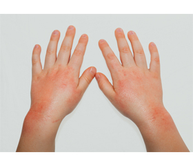
Піт вважається одним з основних факторів загострення АД у всіх вікових групах. Більше того, атопічні екзематозні ушкодження, особливо у дітей, розподіляються переважно в чутливих до поту частинах тіла, таких як ліктьова ямка, підколінна ямка та шия, що означає роль поту в погіршенні атопічних уражень шкіри. Напівочищений антиген поту викликав вивільнення гістаміну з базофілів у 77 % пацієнтів з АД, залежно від специфічного IgE у сироватках пацієнтів з АД. Низка досліджень виявили рівні специфічного IgE проти антигену поту в сироватках хворих на АД на вищому рівні, ніж у контролі, і були пов’язані з тяжкістю АД. Ці спостереження свідчать про те, що пацієнти з АД мають специфічну IgE-опосередковану (тип I) гіперчутливість до вмісту в поті. У дослідженні T. Hiragun та співавт. було визначено білок MGL_1304 Malassezia globosa як основний алерген у людському поту, що індукував вивільнення гістаміну базофілами, отриманими у більшості пацієнтів з АД [44].
Отже, такі чинники, як геном-індуковані варіації, умови навколишнього середовища, спосіб життя, гігієна та імунна система, можуть спричинити порушення шкірних мікробних спільнот, пов’язаних із захворюванням.
Висновки
Захворювання, коли алергія і патоген-асоційовані молекулярні патерни (PAMP) перебувають у тісній взаємодії, — це АД, найбільш поширене запальне захворювання шкіри в усьому світі. Шкіра є найбільш значимим зовнішнім бар’єром і колонізується великою кількістю мікроорганізмів. Пацієнти з АД часто колонізуються та сенсибілізуються умовно-патогенними дріжджами Malassezia. Протигрибкове лікування призводить до поліпшення шкірних симптомів, що може бути наслідком вимирання грибкових, а також через зменшення навантаження на PAMP. Значення дріжджів Malassezia при АД зараз широко вивчається, і потрібні ще дослідження, щоб зрозуміти точну роль цих організмів у перебігу захворювання та загостреннях.
Конфлікт інтересів. Автори заявляють про відсутність конфлікту інтересів та власної фінансової зацікавленості при підготовці даної статті.
Отримано/Received 26.02.2021
Рецензовано/Revised 11.03.2021
Прийнято до друку/Accepted 20.03.2021
Список литературы
1. Cork M.J. et al. Epidermal barrier dysfunction in atopic dermatitis. J. Invest. Dermatol. 2009. 129. 1892-1908.
2. De Benedetto A., Agnihothri R., McGirt L.Y., Bankova L.G., Beck L.A. Atopic dermatitis: a disease caused by innate immune defects? J. Invest. Dermatol. 2009. 129. 14-30.
3. Theelen B., Cafarchia C., Gaitanis G., Bassukas I.D., Boekhout T., Dawson T.L. Jr. Malassezia ecology, pathophysiology, and treatment [published correction appears in Med. Mycol. 2019 Apr 1. 57(3). e2]. Med. Mycol. 2018. 56(1). S10-S25. doi: 10.1093/mmy/myx134.
4. Prohic A., Jovovic Sadikovic T., Krupalija-Fazlic M., Kuskunovic-Vlahovljak S. Malassezia species in healthy skin and in dermatological conditions. Int. J. Dermatol. 2016. 55. 494-504.
5. Iatta R., Cafarchia C., Cuna T. et al. Bloodstream infections by Malassezia and Candida species in critical care patients. Med. Mycol. 2014. 52. 264-269.
6. Velegraki A., Cafarchia C., Gaitanis G., Iatta R., Boekhout T. Malassezia infections in humans and animals: pathophysiology, detection, and treatment. PLoS Pathog. 2015. 11. e1004523.
7. Malassez L. Note sur le champignon du pityriasis simple. Arch. Physiol. 1874. 1. 451-459.
8. Sabouraud R. Maladies du cuir chevelu. II. Les maladies desquamatives. Masson & Cie, Paris, France, 1904.
9. Castellani A., Chalmers A.J. Manual of tropical medicine, 2nd ed. Baillière, Tindal, and Cox, London, United Kingdom, 1913.
10. Faergemann J. Pityrosporum infections. Cutaneous fungal infections. Igaku-Shoin, New York, 1992. 69-83.
11. Gaitanis G., Magiatis P., Hantschke M., Bassukas I.D., Velegraki A. The Malassezia genus in skin and systemic diseases. Clin. Microbiol. Rev. 2012 Jan. 25(1). 106-41. doi: 10.1128/CMR.00021-11. PMID: 22232373; PMCID: PMC3255962.
12. Nagata R., Nagano H., Ogishima D., Nakamura Y., Hiruma M., Sugita T. Transmission of the major skin microbiota, Malassezia, from mother to neonate. Pediatr. Int. 2012. 54. 350-355.
13. Glatz M., Bosshard P.P., Hoetzenecker W., Schmid-Grendelmeier P. The Role of Malassezia spp. in Atopic Dermatitis. J. Clin. Med. 2015 May 29. 4(6). 1217-28. doi: 10.3390/jcm4061217. PMID: 26239555; PMCID: PMC4484996.
14. Saunte D.M.L., Gaitanis G., Hay R.J. Malassezia-Associated Skin Diseases, the Use of Diagnostics and Treatment. Front Cell. Infect. Microbiol. 2020 Mar 20. 10. 112. doi: 10.3389/fcimb.2020.00112. PMID: 32266163; PMCID: PMC7098993.
15. Kolecka A., Khayhan K., Arabatzis M. et al. Efficient identification of Malassezia yeasts by matrix-assisted laser desorption ionization-time of flight mass spectrometry (MALDI-TOF MS). Br. J. Dermatol. 2014. 170. 332-341.
16. Saaf A.M. et al. Global expression profiling in atopic eczema reveals reciprocal expression of inflammatory and lipid genes. PLoS One. 2008. 3. e4017.
17. Brodska P., Panzner P., Pizinger K., Schmid-Grendelmeier P. IgE-mediated sensitization to Malassezia in atopic dermatitis: More common in male patients and in head and neck type. Dermatitis. 2014. 25. 120-126. doi: 10.1097/DER.0000000000000040.
18. Prohic A. Distribution of Malassezia species in seborrhoeic dermatitis: Correlation with patients’ cellular immune status. Mycoses. 2010. 53. 344-349.
19. Gupta A.K. Molecular identification of Malassezia species by amplified fragment length polymorphism (AFLP) and sequence analyses of the internal transcribed spacer (ITS) and large subunit (LSU) regions of ribosomal DNA. J. Am. Acad. Dermatol. 2004. 50. 106.
20. Yim S.M., Kim J.Y., Ko J.H., Lee Y.W., Choe Y.B., Ahn K.J. Molecular analysis of Malassezia microflora on the skin of the patients with atopic dermatitis. Ann. Dermatol. 2010. 22. 41-47. doi: 10.5021/ad.2010.22.1.41.
21. Sandstrom Falk M.H., Tengvall Linder M., Johansson C., Bartosik J., Back O., Sarnhult T., Wahlgren C.F., Scheynius A., Faergemann J. The prevalence of Malassezia yeasts in patients with atopic dermatitis, seborrhoeic dermatitis and healthy controls. Acta Derm. Venereol. 2005. 85. 17-23. doi: 10.1080/00015550410022276.
22. Kaga M., Sugita T., Nishikawa A., Wada Y., Hiruma M., Ikeda S. Molecular analysis of the cutaneous Malassezia microbiota from the skin of patients with atopic dermatitis of different severities. Mycoses. 2011. 54. doi: 10.1111/j.1439-0507.2009.01821.x.
23. Johansson C., Sandstrom M.H., Bartosik J., Sarnhult T., Christiansen J., Zargari A., Back O., Wahlgren C.F., Faergemann J., Scheynius A. et al. Atopy patch test reactions to Malassezia allergens differentiate subgroups of atopic dermatitis patients. Br. J. Dermatol. 2003. 148. 479-488. doi: 10.1046/j.1365-2133.2003.05093.x.
24. Scheynius A., Johansson C., Buentke E., Zargari A., Linder M.T. Atopic eczema/dermatitis syndrome and Malassezia. Int. Arch. Allergy Immunol. 2002. 127. 161-169. doi: 10.1159/000053860.
25. Zargari A., Eshaghi H., Back O., Johansson S., Scheynius A. Serum IgE reactivity to Malassezia furfur extract and recombinant M. furfur allergens in patients with atopic dermatitis. Acta Derm. Venereol. 2001. 81. 418-422. doi: 10.1080/000155501317208363.
26. Johansson C., Eshaghi H., Linder M.T., Jakobson E., Scheynius A. Positive atopy patch test reaction to Malassezia furfur in atopic dermatitis correlates with a T helper 2-like peripheral blood mononuclear cells response. J. Investig. Dermatol. 2002. 118. 1044-1051. doi: 10.1046/j.1523-1747.2002.01758.x.
27. Lange L., Alter N., Keller T., Rietschel E. Sensitization to Malassezia in infants and children with atopic dermatitis: Prevalence and clinical characteristics. Allergy. 2008. 63. 486-487. doi: 10.1111/j.1398-9995.2007.01623.x.
28. Kekki O.M., Scheynius A., Poikonen S., Koskinen A., Kautiainen H., Turjanmaa K. Sensitization to Malassezia in children with atopic dermatitis combined with food allergy. Pediatr. Allergy Immunol. 2013. 24. 244-249. doi: 10.1111/pai.12057.
29. Glatz M., Buchner M., von Bartenwerffer W., Schmid-Grendelmeier P., Worm M., Hedderich J., Folster-Holst R. Malassezia spp.-specific Immunoglobulin E Level is a Marker for Severity of Atopic Dermatitis in Adults. Acta Derm. Venereol. 2015. 95. 191-196. doi: 10.2340/00015555-1864.
30. Sandstrom Falk M.H., Faergemann J. Atopic dermatitis in adults: Does it disappear with age? Acta Derm. Venereol. 2006. 86. 135-139.
31. Ramirez de Knott H.M., McCormick T.S., Kalka K., Skandamis G., Ghannoum M.A., Schluchter M., Cooper K.D., Nedorost S.T. Cutaneous hypersensitivity to Malassezia sympodialis and dust mite in adult atopic dermatitis with a textile pattern. Contact Dermat. 2006. 54. 92-99.
32. Faergemann J. Atopic dermatitis and fungi. Clin. Microbiol. Rev. 2002. 15. 545-563. doi: 10.1128/CMR.15.4.545-563.2002.
33. Gupta A.K., Kohli Y., Summerbell R.C., Faergemann J. Quantitative culture of Malassezia species from different body sites of individuals with or without dermatoses. Med. Mycol. 2004. 39. 243-251. doi: 10.1080/714031025.
34. Findley K., Oh J., Yang J. et al. Topographic diversity of fungal and bacterial communities in human skin. Nature. 2013. 498. 367-370.
35. Wu G., Zhao H., Li C. et al. Genus-wide comparative genomics of Malassezia delineates its phylogeny, physiology, and niche adaptation on human skin. PLoS Genet. 2015. 11. e1005614.
36. Akaza N., Akamatsu H., Sasaki Y. et al. Cutaneous Malassezia microbiota of healthy subjects differ by sex, body part and season. J. Dermatol. 2010. 37. 786-792.
37. Jo J.H., Deming C., Kennedy E.A. et al. Diverse human skin fungal communities in children converge in adulthood. J. Invest. Dermatol. 2016. 136. 2356-2363.
38. Akdis C.A., Akdis M., Bieber T., Bindslev-Jensen C., Boguniewicz M., Eigenmann P., Hamid Q., Kapp A., Leung D.Y., Lipozencic J. et al. Diagnosis and treatment of atopic dermatitis in children and adults: European Academy of Allergology and Clinical Immunology/American Academy of Allergy, Asthma and Immunology/PRACTALL Consensus Report. Allergy. 2006. 61. 969-987. doi: 10.1111/j.1398-9995.2006.01153.x.
39. Zhang E., Tanaka T., Tajima M., Tsuboi R., Kato H., Nishikawa A., Sugita T. Anti-Malassezia-Specific IgE Antibodies Production in Japanese Patients with Head and Neck Atopic Dermatitis: Relationship between the Level of Specific IgE Antibody and the Colonization Frequency of Cutaneous Malassezia Species and Clinical Severity. J. Allergy. 2011. 2011. doi: 10.1155/2011/645670.
40. Selander C., Zargari A., Mollby R., Rasool O., Scheynius A. Higher pH level, corresponding to that on the skin of patients with atopic eczema, stimulates the release of Malassezia sympodialis allergens. Allergy. 2006. 61. 1002-1008. doi: 10.1111/j.1398-9995.2006.01108.x.
41. Balaji H., Heratizadeh A., Wichmann K., Niebuhr M., Crameri R., Scheynius A., Werfel T. Malassezia sympodialis thioredoxin-specific T cells are highly cross-reactive to human thioredoxin in atopic dermatitis. J. Allergy Clin. Immunol. 2011. 128. doi: 10.1016/j.jaci.2011.02.043.
42. Schmid-Grendelmeier P., Fluckiger S., Disch R., Trautmann A., Wuthrich B., Blaser K., Scheynius A., Crameri R. IgE-mediated and T cell-mediated autoimmunity against manganese superoxide dismutase in atopic dermatitis. J. Allergy Clin. Immunol. 2005. 115. 1068-1075. doi: 10.1016/j.jaci.2005.01.065.
43. Yamasaki S., Matsumoto M., Takeuchi O. et al. C-type lectin Mincle is an activating receptor for pathogenic fungus, Malassezia. Proc. Natl. Acad. Sci. USA. 2009. 106(6). 1897-1902. doi: 10.1073/pnas.0805177106,164.
44. Hiragun T., Ishii K., Hiragun M., Suzuki H., Kan T., Mihara S., Yanase Y., Bartels J., Schröder J.M., Hide M. Fungal protein MGL_1304 in sweat is an allergen for atopic dermatitis patients. J. Allergy Clin. Immunol. 2013 Sep. 132(3). 608-615. e4. doi: 10.1016/j.jaci.2013.03.047. Epub 2013 May 29. PMID: 23726042.