Інформація призначена тільки для фахівців сфери охорони здоров'я, осіб,
які мають вищу або середню спеціальну медичну освіту.

Підтвердіть, що Ви є фахівцем у сфері охорони здоров'я.


Всесвітній день боротьби із запальними захворюваннями кишечника
день перший
день другий

Коморбідний ендокринологічний пацієнт

Всесвітній день боротьби із запальними захворюваннями кишечника
день перший
день другий

Коморбідний ендокринологічний пацієнт

Международный эндокринологический журнал Том 17, №3, 2021

Вернуться к номеру

Сироватковий рівень WNT-індукованого протеїну 1 як потенційний біомаркер тиреоїдних вузлів

Авторы: Gulhan Duman, Baris Sariakcali
Division of Endocrinology and Metabolism, Department of Internal Medicine, Cumhuriyet University, Faculty of Medicine, Sivas, Turkey

Рубрики: Эндокринология

Разделы: Клинические исследования

Версия для печати


Резюме

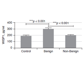
Актуальність. Вузли щитоподібної залози — поширені тиреоїдні захворювання у всьому світі, та їх частота значно зросла за останні десятиліття. Більшість тиреоїдних вузлів зазвичай випадково діагностуються як безсимптомні добро­якісні утворення, виявлені методами візуалізації, проведеними з причин, не пов’язаних із захворюваннями щитоподібної залози. Метою даного дослідження було встановити значення рівня WNT-індукованого протеїну 1 (WISP1) у сироватці крові як допоміжного біомаркера для проведення диференціальної діагностики доброякісних та недоброякісних вузлів щитоподібної залози. Матеріали та методи. У дослідженні брали участь 89 пацієнтів, яким проведено тонкоголкову аспіраційну біопсію, та 43 особи контрольної групи. Серед обстежених жінки становили 72,7 % та 27,3 % — чоловіки. Вони були розподілені на дві групи відповідно до цитологічної оцінки Bethesda: доброякісні (Bethesda 2) та недоброякісні (Bethesda 3–6) утворення. Рівень WISP1 у сироватці крові вимірювали методом імуноферментного аналізу. Результати. У групі з доброякісними вузлами (Bethesda 2) були 58 (43,9 %) пацієнтів, та 31 (23,5 %) — у групі з недоброякісними (Bethesda 3–6) вузлами. Установлено, що розмір утворень був більшим у групі з недоброякісними вузлами, ніж у групі з доброякісними (p = 0,006). Рівень WISP1 у сироватці крові в групі хворих із доброякісними вузлами (Bethesda 2) був вірогідно вищим, ніж у групі з недоброякісними утвореннями (Bethesda 3–6) та осіб конт­рольної групи (p < 0). Різниця між хворими з доброякісними та недоброякісними вузлами відповідно до їх ехогенності була значущою (р < 0,05). У групі з доброякісними вузлами 76,9 % утворень мали змішану ехогенність, 76,7 % — були ізоехогенними, 68,4 % — ізогіпоехогенними та 35,7 % — гіпоехогенними. У хворих із доброякісними вузлами відзначалися найвища гіпоехогенність (64,3 %) і найменша змішана ехогенність (23,1 %). Не встановлено зв’язку між рівнями WISP1 та ехогенністю за допомогою критерію Kruskal-Wallis. Висновки. Згідно з результатами проведеного дослідження, вимірювання WISP1 у сироватці крові дозволяє отримати додаткову інформацію при диференціально-діагностичному аналізі пацієнтів із вузлами щитоподібної залози. Більш високий рівень сироваткового WISP1 дозволяє підтвердити цитологічний діагноз у хворих з доброякісними вузлами щитоподібної залози (Bethesda 2).

Background. Thyroid nodule (TN) is a common thyroid disease worldwide, and it has increased significantly last decades. Most TNs are usually incidental findings of asymptomatic, benign lesions discovered by imaging modalities performed for reasons unrelated to thyroid diseases. The purpose of this study was to investigate the value of serum WNT-induced secreted protein 1 (WISP1) level as a supporting biomarker to perform differential diagnosis of benign and non-benign thyroid nodules. Materials and methods. The study was completed with the 89 patients undergone fine needle aspiration biopsy and 43 controls. The patients were composed of 96 (72.7 %) females and 36 (27.3 %) males. And they were divided into 2 group according to the Bethesda cytological evaluation as Benign (Bethesda 2) and Non-Benign (Bethesda 3–6) groups. Their serum WISP1 levels were measured by an ELISA method. Results. There were 58 (43.9 %) patients in Benign (Bethesda 2) and 31 (23.5 %) in non-Benign (Bethesda 3–6) groups. In the contrary nodule size was bigger in the Non-benign group than that benign group (p = 0.006). The serum WISP1 level in the Benign (Bethesda 2) group was significantly higher than that in the and Non-Benign (Bethesda 3–6) group, and controls (p < 0). The difference between benign and non-benign group accordingly to their echogenicitiy was significant (p < 0.05). In benign group there was 76.9 % mixed echoic nodules, 76.7 % isoechoic nodules 68.4 % isohypoechoic nodules and 35.7 % hypoechoic nodules. In the non-benign group, the highest hypoechoic echo (64.3 %), the least mixed echo (23.1 %), while in the benign group, the most mixed echo (76.9 %), the least hypoechoic echo (35.7 %) was present. There was no relation between WISP1 levels and echogenicity with Kruskal-Wallis H test. Conclusions. According to the preliminary results of current study, addition of serum WISP1 measurement to the differential diagnostic work-up of thyroid nodules patients may provide supportive information. In thyroid nodules patients with Benign (Bethesda 2) category of cytological evaluation, a higher level of serum WISP1 may support cytological diagnosis.


Ключевые слова

вузли щитоподібної залози; ультразвукова діагностика; тонкоголкова аспіраційна біопсія; WISP1

thyroid nodule; thyroid ultrasonography; fine-needle aspiration biopsy; WISP1

Introduction

Thyroid nodule (TN) is a common thyroid disease worldwide, and it has increased significantly last decades [1]. Most TNs are usually incidental findings of asymptomatic, benign lesions discovered by imaging modalities performed for reasons unrelated to thyroid diseases. Thyroid nodules are detected clinically in 5 % of females and in 1 % of males in non-endemic areas. Thyroid malignancy can be detected in up to 15 % of these nodules due to gaining malignancy [2, 3] therefore, the thyroid surgeon is dependent on diagnostic studies to decide when surgery is necessary [4].
In order to further evaluate these nodules, fine-needle aspiration (FNA) is widely accepted as the primary diagnostic tool for the evaluation of TNs owing to its simplicity, safety, and cost-effectiveness [5]. FNA biopsy (FNAB) remains to be mandatory and valuable method used in the evaluation of TNs, but it is still not sufficient as a standard process. Many studies have shown high rate of FNAB specificity (86–100 %) and sensitivity (93–100 %), as well as low rate of false-negative results (3–6 %) [2, 6]. The quality and quantity FNAB applied has been maximized according to the Bethesda cytological evaluation system. A preoperative FNAB should provide all clinical data that can shape the best and appropriate individual treatment for each patient. Yet, the precisely differentiating benign and malignant nodules is crucial and directly related to an accurate management [2, 7].
With technological advancements, diagnostic capabilities of ultrasonography (US) are considerable increased during diagnostic and follow-up procedures as well as guidance during FNAB. Despite the wide application of FNA, the diagnostic yield is limited to 80 to 99 %. Undetermined cytological results always result in confusion and the use of repeated FNA after a non-diagnostic result is still questionable [8, 9]. There is a need new diagnostic tools to reduce unsatisfactory diagnostic results. Within this context, there is a need to the development of a clinically meaningful diagnostic biomarkers to obtain additional clinical data useful for differential diagnosis of TNs without higher cost and increased risk for patients.
Various molecular tests are used to determine whether patients with thyroid nodules that result in indeterminate FNA have cancer. B-Raf proto-oncogene (BRAF), Rat sarcoma viral (RAS) include KRAS, HRAS, NRAS point mutations and ret proto-oncogene (RET/PTC), peroxisome proliferator-activated receptor gamma (PAX8/PPARγ) rearrangements are the most commonly used molecular panels for this purpose. These molecular tests increase the diagnostic power of FNA and thus help to learn more about the biological behavior of the tumor preoperatively [10]. However, no relationship was found between BRAF-V600E KRAS, NRAS mutations in the study conducted by O.İ. Özdamar et al. in thyroid cancer patients with Hashimoto’s disease [11]. 
Because molecular tests are not easy to access, expensive and patients are not willing to use an invasive method such as biopsy, the search for biomarkers that can be easily detected in serum has recently become popular. For this purpose, many biomarkers such as circulating miRNAs, platelets, serum calprotectin, matrix metalloproteinase (MMP), Midkine pleiotropic growth factor, vascular adhesion protein 1 (VAP-1), galectin-3 and interleukins (IL-6, 8, 10) etc. have been investigated to distinguish benign and malign nodules [12].
Up to now, there is no reliable biomarker used to get information about nature of TNs. Among the potential signal transduction growth factors, Wnt inducible signaling pathway protein 1 (WISP1), defined also Cellular Communication Network (CCN4), is a member of the connective tissue growth factor/cysteine-rich 61/nephroblastoma overexpressed family. It is one of the secreted matricellular proteins in the extracellular matrix and has many cellular functions in a highly tissue-specific manner, from cell survival to proliferation. Interestingly, elevated WISP1 expression has also been observed in a variety of cancers such as gastric, colon, lung, liver, and breast cancers and melanoma further confound the role of WISP1 in carcinogenesis [13–15]. 
We hypothesized that WISP1 could be involved in the development of thyroid tumors and investigated the value of serum WISP1 in patients undergone FNAB followed by Bethesda cytological evaluation. As far as we know, no previous study has evaluated the association of serum WISP1 value with Bethesda cytology criteria of TN patients. The objective of this study was to search the value of serum WISP1 level as a supporting biomarker to perform diagnostic work-up of benign and malignant TNs.

Materials and Methods

Patients
The Human Research Ethics Committee of our institution (Registry No: 2019-02/06) approved this study. All patients provided their informed consent before diagnostic procedures. Ultrasound guided FNAB was conducted according to relevant guidelines and regulations. Fine-needle aspiration biopsy accompanied by US was applied to eligible patients with normal TSH who applied to our department for the evaluation of thyroid nodules between January 2019 and July 2019. The patients with diabetes mellitus, obesity (body mass index ≥ 30), chronic kidney disease, chronic heart disease, chronic liver disease, rheumatological disease, cancer and smoking were excluded from the study. The control group included people who applied to find out if they had nodules, but no nodules were detected. The control group also did not have any chronic diseases or habits mentioned above. 
Briefly, FNAB was performed using a 27-gauge needle and a 20-ml syringe under US guidance for all nodules bigger than 1 cm and nodules smaller than or equal to 1 cm with at least one suspicious US finding. FNAB samples were arranged as smears after air-dried.
Cytological evaluation of FNAB specimens was conducted in accordance with the Bethesda System for Reporting Thyroid Cytopathology in 2008 [7]. There are six group according to cytological classification: Bethesda 1, non-diagnostic; Bethesda 2, Benign; Bethesda 3, Atypical of undetermined significance/follicular lesions of undetermined significance; Bethesda 4, Follicular neoplasm/suspicious for follicular neoplasm; Bethesda 5, Suspicious for malignancy; and Bethesda 6, Malignant [16]. In this study, cytology data were presented in the following subsets: Benign (Bethesda 2), non-benign (Bethesda 3–6).
Measurement of WISP1
After overnight fasting, blood samples were collected from all participants into red top tubes (Becton Dickinson, Oxford, UK). Samples were taken on the day of admission to the hospital. The serum sample tubes were allowed to clot before centrifugation. After centrifugation at 4 °C for 15 min at 3.500 rpm, the serum was aliquoted and immediately frozen at –40 °C. 
Serum WISP1 levels measured by Abbkine ELISA kit (China). According to the ELISA kit procedure, the following operations were carried out respectively: standard working solutions were prepared in 5 Eppendorf tubes with concentrations of 480, 240, 120, 60, 30, 15 picograms per milliliter (pg/mL), respectively. 50 µL standards were added to the ELISA plate and 40 µL sample diluent was added to the remaining parts. Ten µl samples were added to the wells with sample diluents. Plate was covered and incubated at 37 °C for 45 min. Plate was washed 5 times with washing solution. Then 50 µL of HRP-conjugate was added to all wells and then it was placed at 37 °C for 30 min. Plate was washed 5 times with washing solution and 50 µl chromogen solution A and 50 µL chromogen solution B was added. It was incubated for 15 min at 37 °C, protected from light. 50 µL of stop solution was added to each well. Absorbance values were read at 450 nm. Concentrations of samples were calculated according to the absorbance values of the standards, the amount of WISP1 was calculated in pg/mL.
Statistical analysis
SPSS 25.0 (IBM SPSS Statistics for Windows, Version 25.0. Armonk, NY: IBM Corp.) package statistics program was used to analyze the data obtained in the study. Before the analysis and evaluations, the compatibility of the obtained data with the normal distribution was tested by performing Histogram and One Sample Kolmogorow-Smirnow test, and it was observed that the data did not have a normal distribution feature. For this reason, besides the frequency distributions, non-parametric tests (Chi-Square, Mann-Whitney U test and Kruskal-Wallis tests) were used. ROC analysis was used to determine the cut-off point to predict benign and non-benign nodules with WISP1 levels. A p value of < 0.05 accepted as statistically significant.

Results

The study was completed with the 89 patients undergone FNAB and 43 controls. The patients were composed of 96 (72.7 %) females and 36 (27.3 %) males. The controls consisted of 28 (65.1 %) females and 15 (34.9 %) males. We divided the patient Benign (Bethesda 2) and Non-Benign (Bethesda 3–6) groups according to the FNAB results. Benign group consisted of 58 (43.9 %) and non-benign group consisted of 31 (23.5 %) patients. There was no difference in terms of age and sex among Benign, non-Benign and Control groups. Selected demographic characteristics of patients undergone FNAB were presented in table 1.
According toWISP1 levels, there is statistically significant difference between benign and non-benign groups. Mean WISP1 level in Benign group is 302.165 ± 88.760, 197.295 ± 48.970 pg/mL in non-Benign group and 234.12 ± 41.39 pg/mL in control group. In the Benign group, the mean level of WISP1 level in women is 286.40 ± 143.73 and 317.93 ± 33.79 pg/mL in men. In the non-Benign group, the average level of WISP1 is 185.81 ± 76.19 in women and 208.78 ± 21.15 pg/mL in men While WISP1 level is higher in benign group, it’s lower in non-benign group (p = 0). This significant difference between benign and non- benign groups is related to the higher WISP1 level in women. In addition, in the ROC analysis, WISP1 cutoff value was found to be 231.24 pg/mL with 73 % sensitivity, 63 % sensitivity and 69 % accuracy respectively. Patients with WISP1 levels above 231.24 pg/mL are more likely to be benign than non-benign. Although WISP1 levels were higher in men in group benign, it was not statistically significant. Mann Whitney U test results used to analyze whether the test results of the patients participating in the study show a significant difference according to the WISP1 levels are given in table 2. Figure 1 depicts the serum WISP1 levels of patients with TNs categorized as Benign (Bethesda 2) and Non-Benign (Bethesda 3–6) groups and controls.
There is a statistically significant difference in Thyroid Stimulating hormone (TSH) levels in patients Benign and non-Benign groups (p < 0.05). Mean TSH level in non-Benign group is 2.5 IU/µIU/mL and 1.9 in Benign group. Accordingly, while TSH level is significantly lower in benign group than in non-benign group (p = 0.048) (table 3). 
There is significant difference in nodule size and nodule count between benign and non-benign groups (p < 0.006). The average nodule size in NonBenign group is 22.92 ± 16.74 mm and 18.57 ± 7.60 mm in Benign group. The size of nodule significantly smaller in benign group than the non-benign one. Similarly, the count of nodules also shows a statistically significant difference between benign and non-benign groups (p < 0.006). While nodule numbers in Benign group is 2.64 ± 1.77, it is 1.92 ± 1.95 in Non-Benign group. The nodule number is higher in benign group than in the non-benign group (table 4). There is no relationship between nodule number and nodule size with WISP1 levels in Sperman Rho test. 
The difference between benign and non-benign group accordingly to their echogenicitiy was significant (p < 0.05). In benign group there was 76.9 % mixed echoic nodules, 76.7 % isoechoic nodules 68.4 % isohypoechoic nodules and 35.7 % hypoechoic nodules. In the non-benign group, the highest hypoechoic echo (64.3 %), the least mixed echo (23.1 %), while in the benign group, the most mixed echo (76.9 %), the least hypoechoic echo (35.7 %) was present (table 5). There was no relation between WISP1 levels and echogenicity with Kruskal-Wallis H test.

Discussion

To the best of our knowledge, current study is the first evaluation of diagnostic value of serum WISP1 in TN patients. The study was completed with the 89 patients undergone FNAB and 43 controls. We divided the patient and groups according to the FNAB results. The patients with Benign (Bethesda 2) category possessed meaningfully higher levels of serum WISP1 compared to that of the controls and Non-Benign (Bethesda 3–6) patients. 
During diagnostic work-up of TNs, with US examination, to determine nature of the TN as benign or malign is not easy in many clinical conditions before decision to surgical intervention. Need for surgical intervention of TN is considerably decreased in last decades after successfully performed FNAB; however, yet it is not clear how to manage long-term follow-up of these patients. One of the important concerns during this follow-up is the possibility of benign to malignant transformation that is a subject of research recently [17]. For shedding light on this subject, Arora et al. conducted a retrospective study investigating the status benign to malignant transformation of thyroid tumors [17]. They noted that the review of our their 10-year experience with thyroid tumors including also screening of some cytological and molecular tumor markers revealed that the rate benign to malignant transformation of thyroid tumors was 2 % in their series.
Generally, FNAB is recommended for TNs of 10 mm or larger in diameter, presenting with a high- or intermediate-suspicion pattern on ultrasonography; TNs 15 mm or larger in diameter, showing a low-suspicion pattern on ultrasonography; and TNs 20 mm or larger in diameter, presenting with a very-low-suspicion pattern on ultrasonography. During first evaluation, TNs considered to be suspicious by medical history or ultrasonographic characteristics should be considered for FNAB when their size is less than 10 mm in diameter [5, 18].
For the appropriate management of patients with TNs, US and FNAB are accepted as the most useful diagnostic modalities. The possibility of malignancy increases significantly with the higher Bethesda rating [19]. Hence, it is useful for the clinicians to be able to predict the possibility of the cytology results. The large-scale use of FNAB in TN patients during surgical evaluation has resulted in an increase in malignant types of specimens as revealed after thyroidectomies by more than 50 %, and additionally, the number of TN patient’s undergone surgery has decreased by 50 % [2].
An identification of malignancy in TNs can reduce missed diagnosis and delayed management, avoiding also surgery for benign TNs, thus decreasing the physical and psychological burden of malignancy. Within this context, since the features of atypical benign and malignant TNs may overlap on routine US [20]. We need new diagnostic modalities to improve our diagnostic capabilities. A relationship with malignancy was found if the level of midline pleotropic growth factor used to distinguish benign and malignant nodules was higher than 323.12 pg/mL, its sensitivity, specificity, and diagnostic accuracy rates of 75.70, 75.00, and 75.31 %, respectively [21]. In another study performed with vascular adhesion protein-1 (VAP-1), the cutoff was found to be 456.6 ng/mL and it was found that the VAP-1 level was lower in patients with thyroid cancer than in healthy individuals without benign nodules with a specificity of 77.4 % and a sensitivity of 66.7 % respectively [22].
Similar to the VAP-1 study mentioned above, we found that WISP1 levels were significantly lower in the non-benign group compared to the benign group in our study. Patients with WISP1 levels above 231.24 pg/mL are more likely to be benign than non-benign with 73 % sensitivity, 63 % sensiti–vity and 69 % accuracy respectively. Therefore, we speculated that if the basal levels of WISP1 levels are known in the benign and non-benign groups, the change of these levels in the future may be a sign of malignant transformation.
It is clear, that evaluation of the thyroid FNABs with the Bethesda cytological evaluation system increases the reliability of cytological diagnosis. However, the impact of Bethesda application may vary among different institutions. Clinicians need to consider the malignancy rate in the Bethesda categories in their hospitals to increase the accuracy of their investigation and decision in the TN patients [23].
According to the results of the study of D.A. Kleiman et al. the authors suggested that decisions about need of FNAB may be based on the presence of suspicious US findings in order to exclude or confirm malignancy [24]. They found that US findings that were significantly associated with malignant diagnosis were microcalcifications, irregular shape, irregular margins and hypoechogenicity of a nodule. They noted that even for smaller nodules of 10 mm with high suspicious pattern, FNAB need to be considered [24]. The US features of TNs in the patients in our study was found somewhat in accordance with those results, although the numbers of patients with these features did not allowed the performance of statistical comparisons.
If the FNAB contains at least six groups that produce ten preserved follicular epithelium cells, accepted as sufficient [7]. Bethesda category 1 cytology results from inadequate sample collection with FNAB and is about 1.8–23.6 % of FNAB results. In many studies, FNAB has shown its sensitivity and specificity to prevent unnecessary surgical procedures, but the technique and the nature of the lesion are very important for an adequate sample. Adequate sample aspiration from sclerotic, calcific and cystic lesions is not an easy procedure [6, 16]. 
We think that adding a biomarker such as serum WISP1 giving information about benign or malign nature of TNs may be helpful to reach a decision about a second FNAB. 
WISP1 gene expression has been shown to be rearranged in diseases such as cancer, diabetic nephropathy, retinopathy and fibrotic diseases. WISP1 is stimulated by the pathway of WINT-1 and B-catenin thus contributes to tumorigenesis [25, 26]. Many studies have demonstrated that WISP1 is related to different kind of tumor development. WISP1 performs its effect using many signal paths, some of these are those mitogen-activated protein kinase (MAPK), protein kinase B (Akt), phosphatidylinositol 3-kinase (PI3K), Jun N-terminal kinase (JNK), caspases, forkhead transcription factors, sirtuins, c-myc oncogene, glycogen synthase kinase 3β (GSK-3β), β-catenin, mammalian target of rapamycin (mTOR) and microRNAs (miRNAs). Through these signal pathways, WISP1 can affect and change the route of cell death programs such as autophagy and apoptosis to protect cytoprotection and tissue repair [27]. 
Excess gene expression of WISP1 regulates cell adhesion of colon cancer and contributes to poor prognosis. WISP1 worsens the course of oral squamous cell carcinoma through angiogenesis. In esophageal squamous cell carcinoma, WISP1 elevation plays a role as a marker of poor prognosis and a regulator of tumor cell radio-sensitivity. These studies vigorously suggest that WISP1 plays an important role in tumor development of various tumors and might be use a target molecule of tumor therapy [28]. WISP1 coordinates cell proliferation, apoptosis, migration, invasion, and chemotherapeutic resistance of glioblastoma [29]. 
High levels of WISP1 are associated with poor prognosis in breast, esophageal and rectal cancers, while in melanoma predicts poor outcome at low WISP1 levels [30–33]. In addition, L.L. Soon et al. demonstrated that overexpression of WISP1 in lung cancer inhibits metastasis, in vitro invasion, and motility through Rac regulatory activation [34]. 
The mouse fibroblast which have over-expressed WISP1, could not form colonies on the soft agar, but when it was injected into the mouse it formed tumors. This suggested that positive paracrine interaction is necessary for tumor development and progression [21]. On the contrary, the fact that H. Shao et al. shown WISP1 is lower in the melanoma cells and fibroblasts activated by melanoma than the fibroblasts in the surrounding tissue suggested the negative paracrine regulatory effect [33]. The crosstalk between tumor cells and the tumor stroma has a considerable effect on tumor progression. Tumor release some growth factors and proteases that will stimulate stromal cells in the surrounding tissues to create a suitable environment for tumor cell progression [35]. This activated carcinoma-associated fibroblasts (CAFs), secrete extracellular matrix proteins and matrix degrading enzymes to regulate microenvironment [35]. 
In colon cancer and in breast cancer WISP1 is upregulated in CAFs more than in fibroblasts at the adjacent normal tissue [36, 37]. In many cancers WISP1 is accumulated in the tumor stroma which around the cancer cells [22, 36–39]. In an experimental animal study, it has been shown that serum and tissue WISP1 levels are high in the early stages of prostate cancer, and the severity of the disease increases with the decrease in tissue and serum WISP1 levels. These studies suggested that WISP1 can be used as a tumor marker such as Prostate Specific Antigen (PSA) [33].
As a result, WISP1 is a dual-acting molecule with an oncogenic effect in some tumors and suppressor in others [40]. Low levels in melanoma increased tumor progression [33], while high levels in lung and prostate cancer suppressed invasion and metastasis [40]. Similar to these cancers, low WISP1 levels were found to be associated with non-benignity in our study.
Although this study has some limitations, whether WISP1, which is used as a follow-up parameter in some malignant cancers, can be used in the follow-up of benign TNs needs to be considered. For this purpose, larger-scale prospective studies are needed to suggest that WISP1 can be used as a diagnostic tool for identifying TNs as benign or malignant. Since recently have revealed a stable and marked rise in the occurrence of thyroid cancer worldwide, the need for biomarkers that may be useful for primary diagnosis and follow-up of TNs in addition to the skilled aspiration, skilled cytological interpretation and rational analysis of cytological and clinical data. 

Conclusions 

Nowadays, transformation mechanism from benign nodule to malignant nodule and affecting factors remain elusive. WISP1 may serve as a potential molecular biomarker for TNs. According to the preliminary results of current study, addition of serum WISP1 measurement to the differential diagnostic work-up of TN patients may provide supportive information. In TN patients with Bethesda 2 category of cytological evaluation, a higher level of serum WISP1 may support cytological diagnosis and long-term follow up. In TN patients with other Bethesda categories of cytological evaluation, a low level of serum WISP1 may support cytological diagnosis. Further studies need to be performed to determine the predictive value of serum WISP1 as a candidate biomarker to differentiate benign and malign TNs according to their final diagnosis after surgery.
Acknowledgement. We would like to thank Assoc. Prof. Halef Okan Dogan for running and evaluating WISP1 Elisa kits.
 
Отримано/Received 02.03.2021
Рецензовано/Revised 25.03.2021
Прийнято до друку/Accepted 05.04.2021

Список литературы

  1. Al Dawish M.A., Robert A.A., Muna A., Eyad A., Al Ghamdi A., Al Hajeri K., Thabet M.A., Braham R. Bethesda System for Reporting Thyroid Cytopathology: A three-year study at a tertiary care referral center in Saudi Arabia. World J. Clin. Oncol. 2017. 8(2). 151-157. doi: 10.5306/wjco.v8.i2.151.
  2. Janczak D., Pawlowski W., Dorobisz T., Janczak D., Dorobisz K., Leśniak M., Ziomek A., Chabowski M. An evaluation of the diagnostic efficacy of fine needle aspiration biopsy in patients operated for a thyroid nodular goiter. Onco Targets Ther. 2016. 9. 5819-5823. doi: 10.2147/OTT.S111275. 
  3. Diana S.D., Hossein G. Epidemiology of thyroid nodules. Best Practice & Research: Clinical Endocrinology & Metabolism 2008. 22(6). 901-911. doi: 10.1016/j.beem.2008.09.019.
  4. Bomeli S.R., LeBeau S.O., Ferris R.L. Evaluation of a thyroid nodule. Otolaryngol Clin. North Am. 2010. 43(2). 229-238. doi: 10.1016/j.otc.2010.01.002.
  5. Haugen B.R., Alexander E.K., Bible K.C., Doherty G.M., Mandel S.J., Nikiforov Y.E., Pacini F. et al. 2015 American Thyroid Association Management Guidelines for Adult Patients with Thyroid Nodules and Differentiated Thyroid Cancer: The American Thyroid Association Guidelines Task Force on Thyroid Nodules and Differentiated Thyroid Cancer. Thyroid. 2016. 26(1). 1-133. doi: 10.1089/thy.2015.0020. 
  6. Yang J., Schnadig V., Logrono R., Wasserman P.G. Fine-needle aspiration of thyroid nodules: a study of 4703 patients with histologic and clinical correlations. Cancer. 2007. 111(5). 306-315. doi: 10.1002/cncr.22955.
  7. Baloch Z.W., LiVolsi V.A., Asa S.L., Rosai J., Merino M.J., Randolph G., Vielh P. et al. Diagnostic terminology and morphologic criteria for cytologic diagnosis of thyroid lesions: a synopsis of the National Cancer Institute Thyroid Fine-Needle Aspiration State of the Science Conference. Diagn. Cytopathol. 2008. 36(6). 425-437. doi: 10.1002/dc.20830. 
  8. Meko J.B., Norton J.A. Large cystic/solid thyroid nodules: a potential false-negative fine-needle aspiration. Surgery. 1995. 118. 996-1003. doi: 10.1016/S0039-6060(05)80105-9.
  9. Shin J.J., Caragacianu D., Randolph G.W. Impact of thyroid nodule size on prevalence and post-test probability of malignancy: a systematic review. Laryngoscope. 2015. 125(1). 263-272. doi: 10.1002/lary.24784. 
  10. Hsiao S.J., Nikiforov Y.E. Molecular approaches to thyroid cancer diagnosis. Endocr. Relat. Cancer. 2014. 21(5). 301-313. doi: 10.1530/ERC-14-0166. 
  11. Özdamar O.İ., Acar G.Ö., Özen F., Zenginkinet T. Assessment of BRAF V600E, KRAS, NRAS and EGFR mutations in Papillary Thyroid Carcinoma and Hashimoto Thyroiditis. Clinical Research. ENT Updates 2020. 10(2). 300-305. doi: 10.32448/entupdates.711666.
  12. Wang W., Chang J., Jia B., Liu J. The Blood Biomarkers of Thyroid Cancer. Cancer Manag. Res. 2020. 12. 5431-5438. doi: 10.2147/CMAR.S261170.
  13. Chiang K.C., Yeh C.N., Chung L.C. et al. WNT-1 inducible signaling pathway protein-1 enhances growth and tumorigenesis in human breast cancer. Sci. Rep. 2015. 5. 8686. doi: 10.1038/srep08686.
  14. Deng W., Fernandez A., McLaughlin S.L., Klinke D.J. 2nd. WNT1-inducible signaling pathway protein 1 (WISP1/CCN4) stimulates melanoma invasion and metastasis by promoting the epithelial-mesenchymal transition. J. Biol. Chem. 2019. 294(14). 5261-5280. doi: 10.1074/jbc. RA118.006122. 
  15. Jia S., Qu T., Feng M. et al. Association of Wnt1-inducible signaling pathway protein-1 with the proliferation, migration and invasion in gastric cancer cells. Tumour Biol. 2017. 39(6). 1010428317699755. doi: 10.1177/1010428317699755.
  16. Evranos B., Polat S.B., Baser H., Ozdemir D., Kilicarslan A., Yalcin A., Ersoy R., Cakir B. Bethesda classification is a valuable guide for fine needle aspiration reports and highly predictive especially for diagnosing aggressive variants of papillary thyroid carcinoma. Cytopathology. 2017. 28(4). 259-267. doi: 10.1111/cyt.12384. 
  17. Arora N., Scognamiglio T., Zhu B., Fahey T.J. 3rd. Do benign thyroid nodules have malignant potential? An evidence-based review. World J. Surg. 2008. 32(7). 1237-1246. doi: 10.1007/s00268-008-9484-1.
  18. Russ G., Bonnema S.J., Erdogan M.F., Durante C., Ngu R., Leenhardt L. European Thyroid Association Guidelines for Ultrasound Malignancy Risk Stratification of Thyroid Nodules in Adults: The EU-TIRADS. Eur. Thyroid J. 2017. 6(5). 225-237. doi: 10.1159/000478927. 
  19. Bongiovanni M., Spitale A., Faquin W.C., Mazzucchelli L., Baloch Z.W. The Bethesda System for Reporting Thyroid Cytopathology: a meta-analysis. Acta Cytol. 2012. 56(4). 333-339. doi: 10.1159/000339959. 
  20. Ha E.J., Baek J.H., Na D.G. Risk Stratification of Thyroid Nodules on Ultrasonography: Current Status and Perspectives. Thyroid. 2017. 27(12). 1463-1468. doi: 10.1089/thy.2016.0654. 
  21. Meng Z., Tan J., Zhang G. et al. Evaluation of serum midkine as a biomarker in differentiated thyroid cancer. Life Sci. 2015. 130. 18-24. doi: 10.1016/j. lfs.2015.02.028.
  22. Hu Z., Zhao P., Zhang K., Zang L., Liao H., Ma W. Evaluation of Serum Vascular Adhesion Protein-1 as a Potential Biomarker in Thyroid Cancer. Int. J. Endocrinol. 2016. 2016. 1-7. doi: 10.1155/2016/6312529.
  23. Edmund S. Cibas and Syed Z. Ali. The 2017 Bethesda System for Reporting Thyroid Cytopathology. Thyroid. 2017. 27(11). doi: 10.1089/thy.2017.0500.
  24. Kleiman D.A., Beninato T., Soni A., Shou Y., Zarnegar R., Fahey T.J. 3rd. Does bethesda category predict aggressive features in malignant thyroid nodules? Ann. Surg. Oncol. 2013. 20(11). 3484-3490. doi: 10.1245/s10434-013-3076-5. 
  25. Pennica D., Swanson T.A., Welsh J.W. et al. WISP genes are members of the connective tissue growth factor family that are up-regulated in wnt-1-transformed cells and aberrantly expressed in human colon tumors. Proc. Natl Acad. Sci USA. 1998. 95(25). 14717-14722. doi: 10.1073/pnas.95.25.14717.
  26. Xu L., Corcoran R.B., Welsh J.W., Pennica D., Levine A.J. WISP1 is a Wnt-1- and beta-catenin-responsive oncogene. Genes. Dev. 2000. 14(5). 585-595. PMID: 10716946.
  27. Kenneth M. WISP1: Clinical Insights for a Proliferative and Restorative Member of the CCN Family. Curr. Neurovasc. Res. 2014. 11(4). 378-389. doi: 10.2174/1567202611666140912115107.142–146. 
  28. Gurbuz I., Chiquet-Ehrismann R. CCN4/WISP1 (WNT1 inducible signaling pathway protein 1): a focus on its role in cancer. Int. J. Biochem. Cell. Biol. 2015. 62. 142-146. doi: 10.1016/j.biocel.2015.03.007. 
  29. Jianghong W., Ziwen L., Hong C., Chunyan D., Xiaowen L. Identification of WISP1 as a novel oncogene in glioblastoma. Oncotarget. 2015. 7(31). 49834-49847. doi: 10.18632/oncotarget.10486.
  30. Taghavi A., Akbari M.E., Hashemi-Bahremani M., Nafissi N., Khalilnezhad A., Poorhosseini S.M., Hashemi-Gorji F., Yassaee V.R. Gene expression profiling of the 8q22-24 position in human breast cancer: TSPYL5, MTDH, ATAD2 and CCNE2 genes are implicated in oncogenesis, while WISP1 and EXT1 genes may predict a risk of metastasis. Oncol. Lett. 2016. 12(5). 3845-3855. doi: 10.3892/ol.2016.5218. 
  31. Nagai Y., Watanabe M., Ishikawa S., Karashima R., Kurashige J., Iwagami S., Iwatsuki M. et al. Clinical significance of Wnt-induced secreted protein-1 (WISP1/CCN4) in esophageal squamous cell carcinoma. Anticancer Res. 2011. 31(3). 991-997. PMID: 21498727.
  32. Davies S.R., Davies M.L., Sanders A., Parr C., Torkington J., Jiang W.G. Differential expression of the CCN family member WISP1, WISP-2 and WISP-3 in human colorectal cancer and the prognostic implications. Int. J. Oncol. 2010. 36. 1129-1136. doi: 10.3892/ijo_00000595.
  33. Shao H., Cai L., Grichnik J.M., Livingstone A.S., Velazquez O.C., Liu Z.J. Activation ofNotch1 signaling in stromal fibroblasts inhibits melanoma growth by upregulating WISP1. Oncogene. 2011. 30. 4316-4326. doi: 10.1038/onc.2011.142.
  34. Soon L.L., Yie T.-A., Shvarts A., Levine A.J., Su F., Tchou-Wong K.-M. Overexpression of WISP1 Down-regulated Motility and Invasion of Lung Cancer Cells through Inhibition of Rac Activation. J. Biol. Chem. 2003. 278(13). 11465-11470. doi: 10.1074/jbc.M210945200.
  35. Mueller M.M., Fusenig N.E. Friends or foes — bipolar effects of the tumour stroma in cancer. Nat. Rev. Cancer. 2004. 4(11). 839-849. doi: 10.1038/nrc1477. PMID: 15516957.
  36. Rupp C., Scherzer M., Rudisch A. et al. IGFBP7, a novel tumor stroma marker, with growth-promoting effects in colon cancer through a paracrine tumorstroma interaction. Oncogene. 2015. 34. 815-825. doi: 10.1038/onc.2014.18.
  37. Bauer M., Su G., Casper C., He R., Rehrauer W., Friedl A. Heterogeneity of gene expression in stromal fibroblasts of human breast carcinomas and normal breast. Oncogene. 2010. 29. 1732-1740. doi: 10.1038/onc.2009.463.
  38. Tanaka S., Sugimachi K., Kameyama T., Maehara S., Shirabe K., Shimada M., Wands J.R., Maehara Y. Human WISP1v, a member of the CCN family, is associated with invasive cholangiocarcinoma. Hepatology. 2003. 37(5). 1122-1129. doi: 10.1053/jhep.2003.50187. 
  39. Ono M., Inkson C.A., Sonn R., Kilts T.M., de Castro L.F., Maeda A., Fisher L.W. et al. WISP1/CCN4: a potential target for inhibiting prostate cancer growth and spread to bone. PLoS One. 2013. 8(8). e71709. doi: 10.1371/journal.pone.0071709. 
  40. Feng M., Jia S. Dual effect of WISP1 in diverse pathological processes. Chinese J. Cancer Res. 2016. 28(6). 553-560. doi: 10.21147/j. issn.1000-9604.2016.06.01.

Вернуться к номеру