Інформація призначена тільки для фахівців сфери охорони здоров'я, осіб,
які мають вищу або середню спеціальну медичну освіту.

Підтвердіть, що Ви є фахівцем у сфері охорони здоров'я.


Всесвітній день боротьби із запальними захворюваннями кишечника
день перший
день другий

Коморбідний ендокринологічний пацієнт

Всесвітній день боротьби із запальними захворюваннями кишечника
день перший
день другий

Коморбідний ендокринологічний пацієнт

Международный эндокринологический журнал Том 17, №1, 2021

Вернуться к номеру

Модель прогнозування тяжкості діабетичної ретинопатії, отримана на основі ендотеліальної дисфункції та маркерів гіпоксії

Авторы: Ya.V. Sirman, I.V. Savytskyi, N.I. Preys
State Institution “Ukrainian Research Institute of Transport Medicine of the Ministry of Health of Ukraine”, Odesa, Ukraine

Рубрики: Эндокринология

Разделы: Клинические исследования

Версия для печати


Резюме

Актуальність. Діабетична ретинопатія — специфічне мікросудинне ускладнення цукрового діабету. Вона залишається провідною причиною втрати зору в працездатного дорослого населення. У пацієнтів із тяжким ступенем діабетичної ретинопатії спостерігається зниження якості життя та погіршення фізичного, емоційного й соціального благополуччя. До того ж на цю когорту пацієнтів витрачається більше ресурсів системи охорони здоров’я. Розробка математичних моделей прогнозування дозволяє більш різнобічно аналізувати експериментальні та клінічні дані. Мета дослідження: розробка моделі прогнозування тяжкості діабетичної ретинопатії на основі маркерів ендотеліальної дисфункції та гіпоксії. Матеріали та методи. Використовували стрептозотоцинову модель цукрового діабету 2-го типу. Визначали рівні фактора Віллебранда, ендотеліну-1, 2,3-дифосфогліцерату, що використовували як змінні. Перехід непроліферативної фази діабетичної ретинопатії в проліферативну на 180-ту добу експерименту підтверджували гістологічно. Результати. Розроблена математична модель для прогнозування досліджуваного патологічного стану на основі біохімічних аналізів крові на ранніх етапах експерименту. Доведена інформативність маркерів ендотеліальної дисфункції, фактора Віллебранда й ендотеліну-1, у прогнозуванні переходу непроліферативної діабетичної ретинопатії в проліферативну. Встановлено значущість визначення рівня 2,3-дифосфогліцерату в еритроцитах разом iз зазначеними вище маркерами функціонального статусу ендотелію на 30-ту добу для прогнозування подальшого перебігу хвороби. Висновки. Доведено значущість показника 2,3-дифосфогліцерату в еритроцитах і зазначених маркерів функціонального статусу ендотелію на 30-ту добу для прогнозування подальшого перебігу досліджуваного патологічного процесу.

Актуальность. Диабетическая ретинопатия — специфическое микрососудистое осложнение сахарного диабета. Она остается ведущей причиной потери зрения у трудоспособного взрослого населения. У пациентов с тяжелой степенью диабетической ретинопатии наблюдается снижение качества жизни и ухудшение физического, эмоционального и социального благополучия. К тому же на эту когорту пациентов затрачивается больше ресурсов системы здравоохранения. Разработка математических моделей прогнозирования позволяет более разносторонне анализировать экспериментальные и клинические данные. Цель исследования: разработка модели прогнозирования тяжести диабетической ретинопатии на основе маркеров эндотелиальной дисфункции и гипоксии. Материалы и методы. Использовали стрептозотоциновую модель сахарного диабета 2-го типа. Определяли уровни фактора Виллебранда, эндотелина-1, 2,3-дифосфоглицерата, которые использовали в качестве переменных. Переход непролиферативной фазы диабетической ретинопатии в пролиферативную на 180-е сутки эксперимента подтверждали гистологически. Результаты. Разработана математическая модель для прогнозирования исследуемого патологического состояния на основе биохимических анализов крови на ранних этапах эксперимента. Доказана информативность маркеров эндотелиальной дисфункции, фактора Виллебранда и эндотелина-1, в прогнозировании перехода непролиферативной диабетической ретинопатии в пролиферативную. Установлена значимость определения уровня 2,3-дифосфоглицерата в эритроцитах вместе с указанными выше маркерами функционального статуса эндотелия на 30-й день для прогнозирования дальнейшего течения болезни. Выводы. Доказана значимость показателя 2,3-дифосфоглицерата в эритроцитах и указанных маркеров функционального статуса эндотелия на 30-й день для прогнозирования дальнейшего течения исследуемого патологического процесса.

Background. According to a wide range of authors, eye damage caused by diabetes mellitus accounts for 80–90 % of the total number of disorders. The development of mathematical predictive models allows for a more versatile analysis of experimental and clinical data. The purpose of the study was to develop a model for predicting the severity of diabetic retinopathy based on a review of markers of endothelial dysfunction and hypoxia. Materials and methods. We used a streptozotocin model of type 2 diabetes mellitus. Determined von Willebrand factor, endothelin-1, 2,3-diphosphoglycerate are used as variables. Also, the transition of the non-proliferative phase of diabetic retinopathy into the proliferative on the 180th day of the experiment was confirmed histologically. Results. We have developed a mathematical model for predicting the studied pathological state based on biochemical blood tests at the early stages of the experiment. We have proven the informative value of endothelial dysfunction markers, von Willebrand factor and endothelin-1, for predicting the transition of non-proliferative diabetic retinopathy into the proliferative phase. We determined the significance of a comprehensive analysis of the level of 2,3-diphosphoglycerate in erythrocytes together with the above markers of the functional status of the endothelium on day 30 for predicting the further course of the disease. Conclusions. We determined the effectiveness of a comprehensive analysis of the level of 2,3-diphosphoglycerate in erythrocytes together with the above markers of the functional status of the endothelium on day 30 for predicting the further course of the pathological process under study.


Ключевые слова

діабетична ретинопатія; прогнозування; математична модель; фактор Віллебранда; ендотелін-1; 2,3-дифосфогліцерат

диабетическая ретинопатия; прогнозирование; математическая модель; фактор Виллебранда; эндотелин-1; 2,3-дифосфоглицерат

diabetic retinopathy; prediction; mathematical model; von Willebrand factor; endothelin-1; 2,3-diphosphoglycerate

Introduction

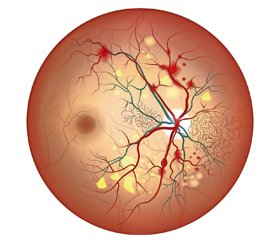
Diabetic retinopathy (DR) is a specific vascular complication of diabetes mellitus (DM) being the major cause of blindness in individuals of active working age in developed economies. According to a wide range of authors, it stands at 80–90 % of total visual disability caused by DM [1, 2].
DR pathogenesis has the following phases: progressive hypoxia that stimulates vascular proliferation and results in adipose degeneration and retinal calcification; microangiopathy of retinal vessels which results in luminal occlusion with hypoperfusion; steal phenomenon with further progression of ischemia that is cause for formation of infiltrates and scars; vascular degeneration with microaneurysms; microinfarctions with exudates, enlargement of proliferating vessels in the retina with shunts and aneurysms; arteriovenous shunts that cause vein dilatation and higher retinal hypoperfusion; retinal detachment due to its ischemic disintegration [3, 4]. The key role of endothelial dysfunction in DR occurrence and progression has been currently proved [5].
Late vascular complications appear to be non-fatal if the prevention and treatment are initiated in time and provided properly [6, 7]. The course of disease should be appropriately predicted to have pathogenic links effectively adjusted at the early stages. Early detection and prediction of microangiopathies and timely influence on modifying risk factors are extremely essential [8].
Pathogenesis of late DM complications has multifactorial nature. Its simplified model includes glycosylation of cell membranes, basal membranes, and proteins. The significant factors are higher vascular permeability, impaired caloric balance, changed metabolism of cell membranes, and induction of polypeptide growth factors. In each particular case, range and severity of certain late complications vary from their paradoxical, almost complete absence, regardless of long duration of the disease, to combination of every possible variant in a severe form [9, 10].
Considering complicated pathogenesis, individual organism specifics, enormous number of factors that differently contribute to pathological growth and closely interact with each other, medication strategy and other impacts on pathological process it seems to be a difficult choice to make. The above shows how difficult is to predict the outcome of disease and need to optimise adjustment of the pathological condition with no experimental study or mathematical method involved. The development of mathematical prediction models enables for a more versatile review of experimental and clinical findings which further contributes to higher efficacy in decision making and individual approach in adjustment of pathological conditions [11].
Considering the above, we have developed the prediction mathematical model of DR severity depending on informative biochemistry markers.
The purpose of the study was to develop a prediction model for severity of diabetic retinopathy based on review of endothelial dysfunction and hypoxia markers.

Materials and methods

Type 2 DM and DR were simulated by means of intraperitoneal administration of streptozotocin (Sigma, USA) diluted in 0.1 M citrate buffer with pH 4.5 [12]. Streptozotocin dose of 55 mg per 1 kg of animal weight was divided into two administrations. Streptozotocin use was preceded by a 28-day high-fat diet [13]. On the 30th day, the levels of von Willebrand factor, endothelin-1, and 2,3-diphosphoglycerate in red blood cells were determined in the blood of this animal group.
The von Willebrand factor was determined with the ristocetin cofactor enzyme-linked immunosorbent assay [14, 15]. Content of serum endothelin-1 as a vasoconstriction marker [16] was evaluated with enzyme-linked immunosorbent assay using DRG reagent sets (Germany). 2,3-diphosphoglycerate in red blood cells is a hypoxia marker [17]. Content of 2,3-diphosphoglycerate was determined with spectrofluorometric method.
A review of transition of non-proliferative phase of diabetic retinopathy into the proliferative one on the 180th day of the experiment was confirmed histologically.
A situation when an explanatory variable in the model takes only two different values tends to occur under study of impact of some subjective and objective factors on presence or absence of some feature in individuals, households, etc. If a study covers n subjects, i.e. if there are n observations, then the fact of presence or absence of such feature in the ith observation may be easily indexed with figures 1 (feature is present) and 0 (feature is not present). Thereby, an indicative (dichotomic, binary) variable y is determined to obtain yi value in the ith observation. And yi = 1 if a feature is present in the ith subject, yi = 0 means a feature is not present in the ith subject [18].
The task of logistic regression is to explain presence or absence of the feature in question with the values (more precisely, combination of values) of some factors (explanation variables). Accordingly, the task includes assessment of parameters of the binary choice model which in general is as follows:
where xi1, …, xip is p value of explanation variables in the ith observation; θ1, …, θp are unknown parameters; εi, …, εn are random errors that show the impact on presence or absence of the investigational feature in the ith subject of any unaccounted additional factors; G(z) is S-like distribution function. We have chosen the standard logistic distribution function (logit-model) to be G(z) function:
We will define a dependent dichotomic variable y as “transition of non-proliferative diabetic retinopathy into the proliferative phase”, i.e. yi = 1 with the fact of transition of non-proliferative diabetic retinopathy into the proliferative phase and yi = 0 with no such fact available.
The following three variable factors were used:
1) x1 as 2,3-diphosphoglycerate in red blood cells;
2) x2 as endothelin-1;
3) x3 as von Willebrand factor.
The protocol was approved by the Ethics Committee of Ukrainian Research Institute of Transport Medicine (protocol 7, 17.09.2019).

Results

Having reviewed a range of studied biochemical blood markers, we determined a relationship between severity of DR and high levels of von Willebrand factor, endothelin-1 (which are common markers for endothelial dysfunction) and 2,3-diphosphoglycerate in red blood cells (hypoxia maker) in the blood of experimental animals.
Let us move to the model analysis. Calculations of the logistic regression parameters and specifications that describe the model adequacy were made in PASW Statistics 18 package. The maximum likelihood estimation was used as loss function; statistical significance of the model was assessed by Chi-square and Hosmer-Lemeshow criteria.
P-value by Hosmer-Lemeshow test is equal to 1 which stands for full consistency of the model with really existing rates in the population sample. This means that the part of dispersion that is explained with logistic regression is equal to 100 %. Additionally, the statistical package gives findings for other criteria, more stable than traditional goodness of fit statistics used in logistic regression, especially to study such small population samples as in our case [19]. The determination unit (determination factor) according to Cox and Snell in our model is equal to 0.632 (63.2 %). This criterion shows the share of impact of all factor features on dispersion of the dependent variable.
Table 1 gives classification of cases and relevant percentage of correct predictions for presence or absence of the fact of transition of non-proliferative DR into the proliferative phase.
Consequently, we have determined that the model derived can by 100 % correctly predict presence or absence of the fact of transition of non-proliferative DR into the proliferative phase.
The following (Table 2) are parameters of the prediction model.
Thus, the logistic regression equation derived is as follows:
To get the relative likelihood for transition of non-proliferative diabetic retinopathy into the proliferative one (within 0–100 %) using the derived logistic regression function, you should multiply the function value G(z) by 100 %.
By inserting parameter values of a certain animal that is not in the studied group in question, we can determine the risk extent for transition of non-proliferative diabetic retinopathy into the proliferative phase.
Here are some examples of how this equation can be used based on particular clinical parameters determined in tested animals which had further confirmed the fact of transition of non-proliferative diabetic retinopathy into the proliferative phase.
Example with high probability for the fact of transition of non-proliferative diabetic retinopathy into the proliferative phase (laboratory animal No 16).
Example with low probability for the fact of transition of non-proliferative diabetic retinopathy into the proliferative phase (laboratory animal No 17).

Discussion

DR is a multifaceted disease with a complex network of metabolic and biochemical alterations that remarkably modify the retinal microenvironment [20]. The research has recently revealed the pivotal role of endothelin in the pathogenesis of diabetic complications, particularly in the regulation of the capillary flow which is affected in retinopathy. Although there are several reviews on various approaches to the treatment of DM, including normalization of glucose and fat metabolism, no reviews in literature have focused on the endothelin system as a therapeutic target or early indicator of diabetic microangiopathy [21].
In fact, novel genetic strategies to earlier detect the upregulation of endothelin-1, or to assess the wide-ranging downstream effects resulted from the activation of the endothelin system in the course of hyperglycemia are a challenging field for new research. Vascular complications of DM can affect not only large and medium arteries resulting in coronary heart disease and peripheral artery diseases but also small vessels leading to retinopathy and nephropathy. Intercellular adhesion molecule-1, vascular cell adhesion molecule-1, E-selectin, and von Willebrand factor are considered as markers of endothelial dysfunction.
By inserting parameter values of a certain animal that is not in the studied group in question, we may determine the risk extent for transition of non-proliferative DR into the proliferative phase.

Conclusions

1. We have proved the role of endothelial dysfunction in growth of investigational diabetic retinopathy.
2. We have developed a mathematical model for prediction of this pathological condition based on biochemical blood tests at early experimental stages.
3. The logistic regression equation derived is as follows:
4. To get the relative likelihood for transition of non-proliferative diabetic retinopathy into the proliferative one (within 0–100 %) using the derived logistic regression function, you should multiply the function value G(z) by 100 %.
5. We have proved the informative value of the endothelial dysfunction markers, von Willebrand factor and endothelin-1, in predicting transition of non-proliferative diabetic retinopathy into the proliferative phase.
6. We have determined the effectiveness of a comprehensive analysis of the level of 2,3-diphosphoglycerate in red blood cells together with the above markers of the endothelial functional status on the 30th day for predicting the further course of the pathological process under study.
Conflicts of interests. Authors declare the absence of any conflicts of interests and their own financial interest that might be construed to influence the results or interpretation of their manuscript.

Список литературы

  1. Wang W., Lo A.C.Y. Diabetic Retinopathy: Pathophysiology and Treatments. Int. J. Mol. Sci. 2018. 19(6). 1816. doi: 10.3390/ijms19061816.
  2. Whitehead M., Wickremasinghe S., Osborne A., Van Wijngaarden P., Martin K.R. Diabetic retinopathy: a complex pathophysiology requiring novel therapeutic strategies. Expert Opin. Biol. Ther. 2018. 18(12). 1257-1270. doi: 10.1080/14712598.2018.1545836.
  3. Bandello F., Lattanzio R., Zucchiatti I., Del Turco C. Pathophysiology and treatment of diabetic retinopathy. Acta Diabetol. 2013. 50(1). 1-20. doi: 10.1007/s00592-012-0449-3.
  4. Tarr J.M., Kaul K., Chopra M., Kohner E.M., Chibber R. Pathophysiology of diabetic retinopathy. ISRN Ophthalmol. 2013. 2013. 343560. doi: 10.1155/2013/343560.
  5. Yau J.W., Rogers S.L., Kawasaki R., Lamoureux E.L., Kowalski J.W., Bek T., Chen S.J. et al. Meta-Analysis for Eye Disease (META-EYE) Study Group. Global prevalence and major risk factors of diabetic retinopathy. Diabetes Care. 2012. 35(3). 556-64. doi: 10.2337/dc11-1909.
  6. Romero-Aroca P., Baget-Bernaldiz M., Pareja-Rios A., Lopez-Galvez M., Navarro-Gil R., Verges R. Diabetic Macular Edema Pathophysiology: Vasogenic versus Inflammatory. J. Diabetes Res. 2016. 2016. 2156273. doi: 10.1155/2016/2156273.
  7. Gonzalez V.H., Campbell J., Holekamp N.M., Kiss S., Loewenstein A., Augustin A.J., Ma J. et al. Early and Long-Term Respon–ses to Anti-Vascular Endothelial Growth Factor Therapy in Diabetic Macular Edema: Analysis of Protocol I Data. Am. J. Ophthalmol. 2016. 172. 72-79. doi: 10.1016/j.ajo.2016.09.012.
  8. Bek T. Diameter changes of retinal vessels in diabetic retinopathy. Curr. Diabetes Rep. 2017. 17. 82. doi: 10.1007/s11892-017-0909-9.
  9. Nentwich M.M., Ulbig M.W. Diabetic retinopathy — ocular complications of diabetes mellitus. World J. Diabetes. 2015. 6(3). 489-99. doi: 10.4239/wjd.v6.i3.489.
  10. Karlberg C., Falk C., Green A., Sjølie A.K., Grauslund J. Proliferative retinopathy predicts nephropathy: a 25-year follow-up study of type 1 diabetic patients. Acta Diabetol. 2012. 49(4). 263-8. doi: 10.1007/s00592-011-0304-y.
  11. Kramer C.K., Retnakaran R. Concordance of retinopathy and nephropathy over time in type 1 diabetes: an analysis of data from the Diabetes Control and Complications Trial. Diabet. Med. 2013. 30(11). 1333-41. doi: 10.1111/dme.12296.
  12. Pasechnikova N.V., Moroz O.A. Protective action of quercetin and lipoate on functional groups of retinal proteins under simulated diabetes. Journal of Ophthalmology. 2015. 3. 76-81. doi: 10.31288/oftalmolzh201537681 (in Russian).
  13. Kaydash O.A., Ivanov V.V., Vengerovsky A.I., Buyko E.E., Schepetkin I.A. The experimental model of type 2 diabetes mellitus caused by a high-fat diet with low-dose streptozotocin in rats. Bulletin of Siberian Medicine. 2020. 19(2). 41-47. doi: 10.20538/1682-0363-2020-2-41-47 (in Russian).
  14. Reinhart K., Bayer O., Brunkhorst F., Meisner M. Markers of endothelial damage in organ dysfunction and sepsis. Crit. Care Med. 2002. 30(5, Suppl). S302-12. doi: 10.1097/00003246-200205001-00021.
  15. He S., Blombäck M., Wallén H., Jeppsson A., Grass S. Global impairments in the haemostasis systems after cardiopulmonary bypass. Thromb. Res. 2017. 151. 63-66. doi: 10.1016/j.thromres.2017.01.006.
  16. Marasciulo F.L., Montagnani M., Potenza M.A. Endothelin-1: the yin and yang on vascular function. Curr. Med. Chem. 2006. 13(14). 1655-65. doi: 10.2174/092986706777441968.
  17. Scott A.V., Nagababu E., Johnson D.J., Kebaish K.M., Lipsitz J.A., Dwyer I.M., Zuckerberg G.S. et al. 2,3-Diphosphoglycerate Concentrations in Autologous Salvaged Versus Stored Red Blood Cells and in Surgical Patients After Transfusion. Anesth. Analg. 2016. 122(3). 616-23. doi: 10.1213/ANE.0000000000001071.
  18. Nosko V.P. Econometrics. Book 2. Part 3, 4. M.: Publishing House “Delo”, 2011. 576 p. (in Russian).
  19. Basilevich K.A., Mazorchuk M.S., Sukhobrus A.A. Modelling in economy, arrangement of production and project management. Information Processing Systems. 2016. 2(139). 149-155 (in Russian).
  20. Sorrentino F.S., Matteini S., Bonifazzi C., Sebastiani A., Parmeggiani F. Diabetic retinopathy and endothelin system: microangiopathy versus endothelial dysfunction. Eye (Lond.). 2018. 32(7). 1157-1163. doi: 10.1038/s41433-018-0032-4.
  21. Siemianowicz K., Francuz T., Gminski J., Telega A., Syzdól M. Endothelium dysfunction markers in patients with diabetic retinopathy. Int. J. Mol. Med. 2005. 15(3). 459-62. PMID: 15702238.

Вернуться к номеру