Introduction
Temporary, or prolonged, extrahepatic cholestasis (EHC), according to many authors, is one of the main etiological factors in the development of cholelithiasis (CL), one of the most common digestive diseases [1–4]. At the same time, if the person does not have a clinical picture of EHC, the diagnosis sounds like CL, chronic cholecystitis, such patients often undergo laparoscopic cholecystectomy, during which bile duct exploration for EHC and its causes is not performed. Complications that develop after laparoscopic cholecystectomy (postcholecystectomy syndrome, mechanical jaundice, chronic pancreatitis, etc.) are frequent causes of diagnostic errors in the subsequent treatment of this group of patients. Despite the development of medical equipment, improvement of surgical skills and many scientific publications, there are many unresolved issues of intraoperative diagnosis of EHC in CL in such individuals. Introduction into clinical practice of ultrasound examination, magnetic resonance cholangiopancreatography (MRCP), endoscopic retrograde cholangiopancreatography (ERCP), endoscopic ultrasonography allow in the vast majority of cases before surgery to establish the cause of bile passage violation in patients with CL in the preoperative period, perform decompression of the biliary tract during surgery, and then either eliminate the cause of bile passage violation with minimally invasive interventions, or prepare the patient for the next stage of surgical treatment [4–9]. However, it is not always possible to determine the cause of EHC in CL using modern diagnostic methods before and during surgery.
At the same time, determining the degree of biliary hypertension is undoubtedly important for the physician, because in the decompensation of bile outflow, the bile hypertension leads to functional and then pathomorphological changes in the hepatobiliary system, which causes a number of complications. Thus, in 1999 E. Corazziari, the head of the group of experts involved in the preparation of criteria for functional disorders of the gallbladder (GB) and sphincter of Oddi (SO), was forced to admit: “We often can’t distinguish between functional disorders of the biliary tract and hidden organic changes. This is due to the peculiarities of the anatomical location of the GB, the imperfection of the research methods used now, and the lack of uniform histological criteria in the assessment of minimal structural changes in the biliary tract” [2]. And even with fully compensated bile outflow, persistent and long-standing biliary hypertension also leads to a number of pathological changes in the biliary tract and liver, causing the transformation of the SO dyskinesia to stenosis, especially in the presence of stones or sludge in the common bile duct [1–6, 10, 11].
The method of cholangiomanometry, proposed in 1868 by Heidenhain, has undergone many modifications over the past century. V. Vinogradov, E. Grishkevich, Mallet-Guy, Lardi, Hessi, Lehner, Roux believed that no bile duct surgery should be done without manometry, and its main purpose — to identify the causes of functional and organic obstructions in the biliary system that disrupt bile flow [10–12].
Therefore, manometry, used in biliary surgery since the mid-nineteenth century and to this day, is the gold standard in the diagnosis of functional disorders of the sphincter apparatus of the biliary system, and the efficiency of method is 92–97 % [2–4, 8].
In 1952, Cannuel, Debray, and Roux Petit proposed that a certain amount of fluid should be passed through the common bile duct over a constant period of time. This method is called debitometry. Several researchers [11, 12] note the great simplicity of this method as an advantage over manometry and more accurate reflection of hydrodynamic changes in pathological conditions of the biliary tract, so the combination of both methods is predicted to increase the possibility of intraoperative diagnosis of functional and organic pathology of the biliary tract with modern methods of visualization of the duct system.
However, to date, the effectiveness of the combined method of manodebitometry in the intraoperative diagnosis of the main causes of EHC in CL has not been established. So, the purpose of our study was to evaluate the effectiveness of the combined method of manodebitometry in intraoperative diagnosis of the causes of different types of extrahepatic cholestasis in complicated cholelithiasis.
Materials and methods
The manodebitometry was performed in 181 patients who were operated at the Department of Surgery of the Digestive Organs of the State Institution “Institute of Gastroenterology of the National Academy of Medical Sciences of Ukraine” for the period from 2013 to 2020. Indications for surgical treatment were: chronic calculous cholecystitis — in 59 (32.6 %) cases; chronic calculous cholecystitis with choledocholithiasis — in 64 (35.36 %); choledocholithiasis after cholecystectomy — in 18 (9.94 %); postcholecystectomy syndrome with stenosing papillitis — in 40 patients (22.1 %).
Patients with the following comorbidities were excluded from the study: viral and autoimmune hepatitis; Caroli disease; Wilson-Konovalov disease; Gilbert’s syndrome; oncological genesis of jaundice; metabolic syndrome.
The age of the examined patients ranged from 27 to 83 years, on average (58.23 ± 1.69) years. Most of the patients were between 40 and 69 years old. There were 52 men (28.73 %) and 129 women (71.27 %).
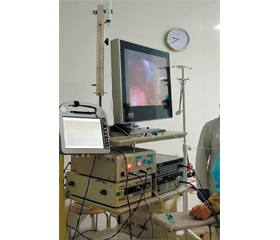
To perform the manodebitometry, we have developed a software and hardware complex consisting of a certified digital domestic manometer MNH-01 ALAN 941118.001, which through a switching cable connected to the invasive pressure sensor Utah DPT-248A on one side, and on the other side — to the three-way valve through a 10-centimeter connector that excludes wetting of the sensor. A 100 ml Janet’s syringe, which serves as a measuring container with a polymer tube with liquid flow regulator from a standard system for intravenous infusion through a compensatory vent cistern, is connected to the second port of the three-way valve. An extension cord is connected to the third port of the valve, which connects to the bile duct catheter. Pressure recording is performed on a tablet computer monitor using the ALAN software package. Data are exchanged between the digital manometer and the computer via a USB port.
The principle of the device is based on converting the displacement of the membrane under the influence of the column of fluid filling the catheter into an electrical signal, which is amplified and digitally converted to a computer, where it is processed numerically, visualized graphically and stored in *.dat format. After cannulation of the choledochus through the GB duct with a drainage tube or retrograde catheter during ERCP, the drainage cannula or catheter in ERCP is connected to an invasive pressure transducer. The sensor is installed at the level of the midaxillary line in the right hypochondrium, which anatomically corresponds to the level of the major duodenal papilla and through the three-way valve connects to a system containing a compensatory vent cistern with a clearly marked fluid level filled with sterile, heated to 37 °C isotonic 0.9% NaCl solution or X-ray contrast solution (Fig. 1).
/42.jpg)
The manodebitometry is a very sensitive method of research, its performance depends on many reasons. The local trauma in the area of the GB, the common bile duct and the major duodenal papilla is of special significance. Careful palpation has a little effect on these parameters, but the allocation of the GB, especially cystic and common bile ducts, is accompanied by the dissection of numerous branches of the hepatic nerve plexus. This leads to a temporary decrease in the SO tonus, a partial decrease in residual pressure, and an increase of fluid debit. Probing or intraductal exploration of the common bile duct and the major duodenal papilla can lead to local edema, spasm of the sphincter, increased residual pressure and decreased debit. Therefore, if manodebitometry is planned, the injury should be as minimal as possible.
To increase the specificity of radiological methods of bile duct visualization [8, 10, 11] and, most importantly, differential diagnosis of the causes of functional and organic SO disorders, we performed intraoperative administration of papillorelaxants (in particular, nitrates or M-cholinolytics), which promote relaxation of the SO. In the absence of a passage of contrast agent through the SO, we performed the SO relaxation test — intravenous bolus administration of 10–20 mg (1–2 ml) of isosorbide dinitrate or 20 mg of hyoscine butylbromide, and then after 2 minutes the study was repeated.
Thus, the SO relaxation test was considered positive if the passage of contrast fluid restored after the introduction of papillorelaxants, and negative — if it did not. Due to non-specificity of symptoms, when they are detected, neoplasms of the pancreatobiliary zone and choledocholithiasis should be excluded in the first place.
After filling the system, the compensating cistern and the choledochus, the initial pressure parameters in the duct were registered. The pressure required for manodebitometry was created by further increasing the liquid column until the pressure values were registered on a digital manometer with a fixed set level of 300 mm of water column (mm H2O) and with the appearance of relative isoline on a portable tablet computer, after which the manodebitometric camera was fixed on a tripod. Then we performed registration of pressure and debit in accordance with physiological norms after V. Vinogradov (1964) [11].
The concentration of total bilirubin in the serum was determined according to the instructions to the ELITech kits (France). The activity of serum alanine aminotransferase (ALT), aspartate aminotransferase (AST), alkaline phosphatase (AP), γ-glutamyltransferase (GGT) was evaluated by ultraviolet kinetics, recommended by the International Federation of Clinical Chemistry and Laboratory Medicine, according to the instructions of the ELITech kits (France). The presence of endogenous intoxication was determined by the content of middle molecular weight peptides (MMP) according to V.V. Nikolaychuk. The MMP fraction consists of aromatic amino acids, which are part of proteins, collagen fibers, aromatic amino acids, among which tyrosine and tryptophan occupy a significant place, and therefore an increase in the serum content of MMP is a marker of activation of catabolic processes in the body. Fibrotic processes were evaluated by the content of free hydroxyproline and glycosaminoglycans. In serum, the content of glycosaminoglycans was determined according to Remington, free hydroxyproline — by Osadchuk, the activation of the inflammatory process in patients showed a change in the level of alpha-1-acid glycoprotein, the content of which was evaluated by Weimer. Biochemical parameters were assessed according to their content in blood of relatively healthy 20 people (control group).
The ultrasound examination was performed in all patients, and if choledocalculosis is suspected, ERCP was carried out in 79 (43.64 %) cases as diagnostic and treatment manipulation, and MRCP — in 58 (32.04 %) people.
For the assessment of the motoric and evacuation function of the GB before the operation in 52 (28.72 %) patients who have no contraindication, such as small stones or inhomogeneous content in the GB or choledohus on ultrasound, the following method was used: the initial volume (V1) of the GB was determined on an empty stomach, then after a cholekinetic breakfast (20 g of sorbitol dissolved in 50 ml of warm water), the volume of the GB was determined per 1 minute for the first 10 min and then every 10 min until it relaxed. The volume of the GB after its maximum contraction (V2) was used to determine the efficiency of bile secretion (EBS):
The EBS was considered normal if 20–40 minutes after a cholekinetic breakfast, its maximum was 40–70 % of the initial volume of the GB. Assessment of the functional state of the GB was performed taking into account the primary reaction, latency period, and the time of maximum contraction of the GB [13].
During the operation, all patients underwent manodebitometry, the indicators of which were compared with the data of X-ray cholangioscopy and cholangiography. The process of cannulation of the cystic duct took most time for manodebitometry, and in fact, the study time, depending on the need for repeated measurements, averaged (7.2 ± 3.6) minutes.
The research was performed in compliance with the rules of ethical principles of scientific medical research with human participation, approved by the Declaration of Helsinki (1964–2013), ICH GCP (1996), EEU Directive No. 609 (dated 24.11.1986), orders of the Ministry of Health of Ukraine No. 690 dated 23.09.2009, No. 944 dated 14.12.2009, No. 616 dated 03.08.2012. Each patient signed an informed consent to participate in the study, with all measures to ensure the anonymity.
Statistical analysis of the obtained data was performed using Microsoft Excel 2010 and Statistica 12.0. The median (Me), lower and upper quartiles (Q25; Q75) were used to describe the data. The comparisons were performed by means of a nonparametric criterion (Mann-Whitney U test). Mean values were compared using Student’s t-test. The difference was considered significant if the achieved significance level (p) was less than 0.05. To assess the diagnostic effectiveness of the indicators, receiver operating characteristic (ROC) analysis was used to determine the area under the ROC-curve (AUC), the optimal threshold value, sensitivity, and specificity.
Results
In 30 patients diagnosed with chronic calculous cholecystitis, in the preoperative period according to biochemical studies, blood markers of cholestasis were not detected (control group), but they had indirect clinical signs of EHC in the past medical history: episodes of scleral yellowing, urine darkening. Also, these individuals had a history of EHC predictors, namely: pregnancy, oral contraceptives or other hormonal drugs intake, diabetes, episodes of prolonged starvation or parenteral nutrition, truncal vagotomy; therefore, they were allocated to a separate “temporary” type 0 group of EHC (with anamnestic episodes of extrahepatic cholestasis).
By the content of biochemical markers, 4 types of EHC were identified among 151 patients:
a) type I — without jaundice and without damage to hepatocytes (n = 50): the average level of bilirubin was (23.5 ± 1.7) μmol/l, ALT — (38.20 ± 3.45) IU/l, AST — (36.90 ± 4.14) U/l, which indicated the absence of cytolysis, and AP of (284.0 ± 32.4) U/l and GGT of (247.0 ± 34.6) U/l — the presence of EHC; the level of MMP was (726 ± 36) g/l;
b) type II — without jaundice with hepatocyte damage (n = 38): the average level of bilirubin was (34.7 ± 3.2) μmol/l, ALT — (136.0 ± 27.8) IU/l, AST — (84.13 ± 15.50) U/l, which indicated the presence of cytolysis, and AP of (62.0 ± 32.8) IU/l and GGT of (345.0 ± 37.6) IU/l — the presence of EHC; the level of MMP was (902.0 ± 56.4) g/l;
c) type III — with jaundice without damage to hepatocytes (n = 36): the average level of bilirubin was (159.00 ± 12.75) μmol/l, ALT — (39.8 ± 5.1) IU/l, AST — (40.4 ± 6.8) U/l, which indicated the presence of cytolysis, and AP of (310.0 ± 59.1) IU/l and GGT of (260.0 ± 41.4) IU/l — the presence of EHC; the level of MMP was (1,050.0 ± 75.7) g/l;
d) type IV — with jaundice and hepatocyte damage (n = 27): the average level of bilirubin was (313.8 ± 28.1) μmol/l, ALT — (287.0 ± 44.6) IU/l, AST — (242.00 ± 49.67) U/l, which indicated the presence of cytolysis, and AP of (630.0 ± 81.2) IU/l and GGT of (610.0 ± 69.9) IU/l — the presence of EHC; the level of MMP was (1,139.0 ± 78.4) g/l.
Indicators of the cause of EHC in complicated CL, which were established before and clarified during surgery by radiological studies, were compared with the parameters of manodebitometry (Table 1).
Comparison of manodebitometry data with the indicators of the causes of EHC in complicated CL, established using ultrasound before and refined radiologically during the operation, allowed determining that in the form of manodebitometric coefficient, parameters of the main causes of each type of EHC differ statistically and reflect the staged development of EHC in complicated CL.
Discussion
Preoperatively undiagnosed phenomena of isolated stenosing papillitis at different stages of formation were detected intraoperatively using manodebitometry with radiological verification in 34 (18.78 %) patients, including:
— 13 (7.18 %) people with type I of EHC;
— 19 (10.49 %) individuals with type II of EHC, including 7 (3.86 %) cases of combination with choledocholithiasis;
— 8 (4.41 %) patients with type III of EHC, including 3 (1.65 %) cases of combination with choledocholithiasis;
— 7 (3.86 %) people with type IV of EHC, of which in 3 (1.65 %) cases it was combined with choledocholithiasis.
The combination of choledocholithiasis and stenosing papillitis was found in 13 (7.18 %) patients.
Isolated SO dyskinesia as functional disorder due to laparoscopic cholecystectomy in the past medical history was detected in 6 (3.31 %) people with type 0 of EHC and in 7 (3.86 %) — with type III of EHC.
In 25 patients (3 (1.65 %) — with type 0 of EHC and 22 (12.15 %) — with type II of EHC), the phenomena of the SO dysfunction were determined in combination with structural disorders at the stage of formation (SO dyskinesia + stenosing papillitis at the stage of formation, degree 0–1).
It should be noted that patients with type 0 of EHC at the time of surgery did not have any biochemical predictors of cholestasis.
The diagnosed stage of stenosing papillitis formation corresponded to a certain type of EHC; however, according to the results of studies, 11 patients with type II of EHC (6.07 % of the total number and 28.94 % of those with type II of EHC) had the phenomena of stenosing papillitis, manometric parameters of which does not clearly correspond to the existing classification (V. Vinogradov, E. Grishkevich, 1963), so those cases were defined as the SO stenosis degree I–II.
Due to the certain variability of the parameters of the residual pressure of choledochus and fluid debit at the beginning of the study and after the injection of papillorelaxants (nitrates, M-cholinolytics) at the end of the study, we decided to calculate an integral numerical coefficient:
For example, in patients with type 0 of EHC in whom SO dysfunction was detected, the initial pressure and debit were 140.6 mm H2O and 7.4 ml/min, final ones — 108 mm H2O and 27 ml/min, respectively (against the background of the introduction of papillorelaxants after the first minute of the study, Fig. 2), while T = 1.3 (140.6/108), D = 0.27 (7.4/27), and C = 1.57 (1.3 + 0.27).
According to the ratio of the sum of the values of the pressure fraction at the beginning and end of the study and the fraction of fluid debit during one minute and at the end of the study (before and after the relaxation test), the following coefficients can be distinguished for intraoperative pathology screening (Fig. 3).
Therefore, as can be seen from the distribution, the determined coefficient for different causes of cholestasis in organic disorders of the SO will always be in the range of 1.8–2.03, and in functional disorders — always lower than 1.8. The coefficient of uncomplicated chronic calculous cholecystitis is in the range of 2.35–2.6, and an index above 2.6 allows us to suspect the presence of floating stones.
Thus, the use of a combined method of manodebitometry in the intraoperative diagnosis of the main causes of different types of EHC in complicated CL allowed in 34 (18.78 %) patients to diagnose organic disorders as the cause of EHC and in 38 (20.99 %) — functional disorders of the sphincter apparatus. The number of intraoperatively detected structural and functional disorders is 39.77 % among those who underwent surgery.
When conducting ROC-analysis, the use of a combined method of manodebitometry as a diagnostic screening for functional and organic causes of EHC in complicated CL allowed establishing a high quality of diagnostic model using a coefficient as the optimal classification threshold (Fig. 4) with an accuracy of 88.9 %, sensitivity 93.6 %, specificity 80 %, as AUC was 0.9311 (95% confidence interval 0.918–0.929; p < 0.0001).
Therefore, the analysis of the results of evaluating the use of the combined method of manodebitometry in the examined patients showed high sensitivity and specificity of the obtained data in the established organic and functional EHC in complicated CL.
Conclusions
1. The combined method of manodebitometry, which consists in determining the pressure in the extrahepatic bile ducts and the debit of perfused fluid through the terminal part of choledochus per unit time, allows increasing up to 34.8 % the detection of EHC in complicated CL and establishing its functional and organic causes during surgery.
2. Due to its simplicity, easy reproducibility and diagnostic value (accuracy 88.9 %, sensitivity 93.6 %, specificity 80 %), manodebitometric study according to the developed method can be recommended for practical application for the recognition of EHC type in complicated CL, its main causes and intraoperative determination of indications for their correction during surgery.
3. The use of manodebitometric examination of the choledochus with a pharmacological test for relaxation allows forming a clear idea of the functional state of the muscular apparatus of the sphincter of Oddi and detecting its dysfunction.
4. Manodebitometry is considered necessary in all doubtful cases, which can significantly reduce the number of undiagnosed causes of EHC in complicated CL and, in the long run, the development of postoperative complications.
Conflict of interest. The authors declare the absence of conflict of interest and their own financial interest in the preparation of this article.

/42_2.jpg)
/43.jpg)
/45_2.jpg)
/44.jpg)
/45.jpg)