Інформація призначена тільки для фахівців сфери охорони здоров'я, осіб,
які мають вищу або середню спеціальну медичну освіту.

Підтвердіть, що Ви є фахівцем у сфері охорони здоров'я.


Всесвітній день боротьби із запальними захворюваннями кишечника
день перший
день другий

Коморбідний ендокринологічний пацієнт

Всесвітній день боротьби із запальними захворюваннями кишечника
день перший
день другий

Коморбідний ендокринологічний пацієнт

Международный эндокринологический журнал Том 16, №4, 2020

Вернуться к номеру

Impact of compliance to a gluten-free diet on vitamin and trace element deficiencies in celiac patients

Авторы: Hasret Ayyildiz Civan
Bakirkoy Dr. Sadi Konuk Training and Research Hospital, Istanbul, Turkey

Рубрики: Эндокринология

Разделы: Клинические исследования

Версия для печати


Резюме

Актуальність. Целіакія — хронічне захворювання імунного генезу, що характеризується затримкою росту та маль­абсорбцією, пов’язаною з пошкодженням слизової оболонки і запаленням тонкої кишки у генетично схильних людей внаслідок впливу глютену. При лікуванні целіакії досягається клінічне, гістологічне та серологічне поліпшення стану шляхом дотримання безглютенової дієти. Мета дослідження: оцінити рівень вітамінів та мікроелементів у хворих на целіакію за умов дотримання безглютенової дієти. Матеріали та методи. Під спостереженням перебували 77 пацієнтів з діагнозом «целіакія», виявленим ретроспективно. Усі випадки були підтверджені гістологічно за класифікацією Marsh, усі па­цієнти обстежені на тлі дотримання безглютенової дієти. Демографічні особливості, вік початку захворювання, результати клінічного обстеження, антропометричні показники та лабораторні результати порівнювали у групах хворих, які дотримувалися дієти, та за її відсутності. Результати. До дослідження були включені 77 хворих (48 жінок і 29 чоловіків) із целіакією, середній вік яких становив 9,81 ± 4,73 року. Переважно у пацієнтів виявлені гістологічно за класифікацією Marsh типи 3a (n = 22) і 3b (n = 20). Результати серологічного скринінгу показали, що у 40,3 % випадків (n = 31) хворі дотримувалися безглютенової дієти, тоді як у 59,7 % (n = 46) — не дотримувалися. У хворих цієї групи відзначалися вірогідно нижчі показники вмісту вітаміну В12, вітаміну D, фолатів, цинку та селену порівняно з групою хворих, які дотримувалися відповідної дієти (p = 0,000; 0,000; 0,000; 0,000 та 0,031 відповідно). Крім того, був виявлений вірогідно вищий середній рівень загального IgA в сироватці крові у групі хворих без дотримання дієти порівняно з групою хворих, які дотримувалися безглютенової дієти (p = 0,027). Висновки. Встановлена висока ефективність безглютенової дієти для корекції недостатності та дефіциту вітамінів і мікроелементів. Необхідно детально інформувати пацієнтів з целіакією та їх родини про неухильне пожиттєве дотримання безглютенової дієти, хоча дотримання такого харчування створює чимало соціальних і практичних труднощів.

Актуальность. Целиакия — хроническое заболевание иммунного генеза, характеризующееся задержкой роста и мальабсорбцией, связанной с повреждением слизистой оболочки и воспалением тонкой кишки у генетически предрасположенных людей в результате воздействия глютена. При лечении целиакии достигается клиническое, гистологическое и серологическое улучшение состояния путем соблюдения безглютеновой диеты. Цель исследования: оценить уровень витаминов и микроэлементов у больных целиакией при соблюдении безглютеновой диеты. Материалы и методы. Под наблюдением находились 77 пациентов с диагнозом «целиакия», выявленным ретроспективно. Все случаи были подтверждены гистологически по классификации Marsh, все пациенты обследованы на фоне соблюдения безглютеновой диеты. Демографические особенности, возраст начала заболевания, результаты клинического обследования, антропометрические показатели и лабораторные результаты сравнивали в группах больных, которые придерживались диеты, и при ее отсутствии. Результаты. В исследование были включены 77 больных (48 женщин и 29 мужчин) с целиакией, средний возраст которых составил 9,81 ± 4,73 года. В основном у пациентов обнаружены гистологически по классификации Marsh типы 3a (n = 22) и 3b (n = 20). Результаты серологического скрининга показали, что в 40,3 % случаев (n = 31) больные соблюдали безглютеновую диету, тогда как в 59,7 % (n = 46) — не соблюдали. У больных этой группы отмечались достоверно более низкие показатели содержания витамина В12, витамина D, фолатов, цинка и селена по сравнению с группой больных, которые соблюдали соответствующую диету (p = 0,000; 0,000; 0,000; 0,000 и 0,031 соответственно). Кроме того, был обнаружен достоверно более высокий средний уровень общего IgA в сыворотке крови в группе больных без соблюдения диеты по сравнению с группой больных, которые соблюдали безглютеновую диету (p = 0,027). Выводы. Установлена высокая эффективность безглютеновой диеты для коррекции недостаточности и дефицита витаминов и микроэлементов. Необходимо детально информировать пациентов с целиакией и их семьи о неуклонном пожизненном соблюдении безглютеновой диеты, хотя соблюдение такого питания создает немало социальных и практических трудностей.

Background. Celiac disease (CD) is a chronic immune-mediated disorder characterized by growth retardation and malabsorbtion related to mucosal damage and inflammation of small intestine in genetically predisposed people as a result of gluten exposure. In CD treatment, clinical, histological and serological improvement is possible with gluten-free diet. Thus, we aimed to assess the vitamin and trace element levels of CD patients in regard to their compliance with gluten-free diet. Materials and methods. In our study, 77 patients diagnosed with CD were evaluated retrospectively. All individuals were assessed with Marsh classification histopathologically and surveyed when they follow a gluten-free diet. Demographic features, age of disease onset, physical examination findings, anthropometric measurements and laboratory findings along with clinical and laboratory outcomes of patients after a gluten-free diet were compared between compliant to diet and non-compliant to diet groups. Results. A total of 77 individuals, 48 females and 29 males with a diagnosis of CD and mean age of 9.81 ± 4.73 years on admission, were reqruited in our study. Patients were mostly found to have Marsh type 3a (n = 22) and Marsh type 3b (n = 20) histopathologically. The results of serological screening revealed that 40.3 % of people (n = 31) were compliant with diet whereas 59.7 % (n = 46) — non-compliant. Non-compliant group had significantly lower mean vitamin B12, vitamin D, folate, zinc and selenium levels compared to compliant group (p = 0.000; 0.000; 0.000; 0.000 and 0.031, respectively). In addition, a significantly higher mean serum total IgA level was detected in non-compliant group in comparison to compliant group (p = 0.027). Conclusions. High efficacy of gluten-free diet in correcting nutritional insufficiencies and deficiencies was shown. Thus, there is no doubt that informing patients and their families about lifelong gluten-free diet in detail is beneficial although this treatment contains many social and practical difficulties.


Ключевые слова

целіакія; безглютенова дієта; мальабсорбція; вітамін D; вітамін В12

целиакия; безглютеновая диета; мальабсорбция; витамин D; витамин В12

celiac disease; gluten-free diet; gluten; malabsorption; vitamin D; vitamin B12

Introduction

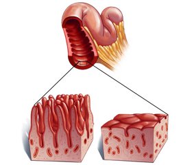
Celiac disease (CD) is a chronic immune-mediated disorder characterized by malabsorbtion and growth retardation in children as a result of inflammation, infiltration and villous atrophy in small intestine [1]. There is a lifelong intolerance to gluten of wheat in particular, together with grain proteins of rye, barley and sometimes contaminated oat products in genetically predisposed people [2]. 
Even if the disease presents often with chronic diarrhea, weight loss and bloating, asymptomatic course is also likely. Moreover, a broad spectrum of clinical symptoms including stomach ache and chronic fatigue are found along with disorders related to vitamin and mineral uptake such as iron deficiency anemia, vitamin D deficiency and calcium deficiency. Although the prevalence of CD is reported to be almost 1 % in Europe, actual prevalence is considered as high as 7–10 fold due to its insidious presentation and globally increased incidence in recent years [2–4].
Though histopathological assessment is regarded as the gold standard of diagnosis, tissue transglutaminase antibody, anti-endomysial antibody and human leukocyte antigene tests are among first line diagnostic tools [5]. In fact, there is a well known pathophysiological interaction between environmental and genetic risk factors in CD. Accordingly, CD does not develop in absence of allelle coding HLA-DQ2 or HLA-DQ8 proteins while presence of that allelle does not always suffice for the disease to emerge. In addition, a mild gluten exposure of celiac patients may lead to mucosal damage, however clinical, histological or even serological (TGA/G) improvement is likely after a gluten-free diet [1, 2]. 
Thus, we purposed to analyze the vitamin and trace element levels of patients we followed up with a diagnosis of CD, in regard to dietary compliance after a gluten free diet GFD) therapy.

Materials and methods

This study was conducted in pediatric gastroenterology outpatient clinics of Istanbul Training and Research Hospital between January 2017 and June 2018 in accordance with the local ethics committee approval of 07.01.2019/201-01-24. 
A total of 77 patients being between 1 to 18 years of age diagnosed with CD who have no other chronic illness leading to additional malabsorbtion were investigated retrospectively. Demographic features of patients, age of disease onset, physical examination findings, anthropometric measurements and laboratory findings along with clinical and laboratory outcomes of patients after a GFD were compared between compliant to diet and non-compliant to diet groups. Biochemical analysis of serum derived from patients was performed with the Abbott Architect c8000 autoanalyzer (Abbott Diagnostics, Illinois, USA) by using electro-chemiluminescence method whereas serum 25-hydroxyvitamin D3, vitamin B12 and folate levels were measured and reported by using test kits compatible with chemiluminescence method in Roche Cobas 6000 (Tokyo, Japan) immunological autoanalyzer device. 
Serological screening for CD was carried out with enzyme-linked immunosorbent assay by using anti tissue transglutaminase antibodies (TGAb). All patients were also screened with immunoflorescence method ((INOVA, San Diego, Calif., USA) for anti-endomysial antibodies, umbilical cord and fluorescein isothiocyanate conjugated anti-human IgA. To detect serum IgA deficiency, total serum IgA levels were assessed with nephelometric method. In addition, presence of HLA antigenes (DQ2 and DQ8) were investigated by using polimerase chain reaction (PCR) technique on genomic DNA isolated from peripheral blood of all patients. Patients with positive TGA and EMA antibodies got a definite diagnosis after the endoscopic biopsy taken from distal duodenum and cases were then evaluated histopathologically in regard to Marsh classification. All CD cases were followed up with gluten-free diet. 
Statistical analysis. For the statistical analysis of our study, the 21.0 version of SPSS software ((Statistical Package of the Social Sciences, IBM, Armonk, NY, USA) was utilized. Descriptive statistics were expressed as mean ± standard deviation or median (minimum-maximum) for discrete and continuous quantitative variables whereas categorical variables were given as number of cases and percentage (%). For the comparison of categorical variables, cross table statistics were used (Chi square, Fisher). While normally distributed parametric data sets were compared by using Student t-test and ANOVA, non-parametric data with abnormal distribution were compared via Mann Whitney U and Kruskal Wallis tests. Comparisons between multiple groups were carried out with post hoc Tukey analysis. In results, p < 0.05 was defined as statistically significant. 

Results

A total of 77 cases consisting of 48 females (62.3 %) and 29 males (37.7 %) followed up with the diagnosis of CD were reqruited in our study. Female to male ratio of patients was found to be 1.65 and mean age of all patients on admission was 9.81 ± 4.73 (range of distribution; 2–19) years. In particular, mean age of males was 9.25 ± 4.23 years whereas mean age of females was found to be 10.16 ± 5.02 years (p = 0.381). Additionally, overall mean age of cases at diagnosis was 2.28 ± 2.37 years (range of distribution: 0–11years) (Table 1). A total of 57 patients from our study population who underwent gastroduedonoscopy and intestinal biopsy were investigated histopathologically which revealed that 22 of those had Marsh type 3a, 20 Marsh type 3b, 13 Marsh type 3c and two Marsh type 1 and 2 disease.
As a result of serological screening in our study carried out upon TG IgA, TG IgG, EMA-IgA and EMA-IgG levels, 40.3 % of cases (n = 31) were compliant to diet and the remaining 59.7 % (n = 46) were non-compliant. In the non-compliant group, mean serum IgA level was 137.94 ± 66.02 mg/dl, vitamin B12 level 328.43 ± 167.22 pg/ml, vitamin D level 21.70 ± 13.38 ng/ml, folate 9.46 ± 5.37 mg/l, zinc 70,61 ± 11,25 µg/dl and selenium 42.83 ± 8.47 µg/dl. Conversely, mean serum total IgA level was found to be 109.55 ± 64.17 mg/dl, vitamin B12 level 496.71 ± 179.94 pg/ml, vitamin D 31.24 ± 10.90 ng/ml, folate 15.03 ± 5.08 mg/l, zinc 82.32 ± 9.84 µg/dl and selenium 49.91 ± 5.77 µg/dl in the group compliant to diet. Hence significantly higher mean serum total IgA levels were observed in non-compliant group compared to compliant group (p = 0.027). In addition, significantly lower mean serum vitamin B12, vitamin D, folate, zinc and selenium levels were noted in non-compliant group compared to compliant group (p values; 0.000; 0.000; 0.000; 0.000 and 0.031, respectively).

Discussion

CD developed into a crucial global health issue as its prevalence increases in recent years and its atypical and asymptomatic presentations suggest presence of many undiagnosed cases. Accordingly, current data show that children and adolescents with a higher mean age compared to that of past reports are diagnosed with CD [6]. Predominance of female gender in CD was also reported many times [7]. N.R. Reilly et al. reported a mean age of 8.3 years based on 318 pediatric patients of whom 57 % were female and diagnosed with CD histopathologically [8]. E. Lurz et al. also reported a group of 94 pediatric patients at a mean age of 6.8 years diagnosed with CD, which displayed a female predominance of 62 % [9]. Consistent with the published data, female/male ratio in our study was calculated as 1.65 and the mean age on admission was found to be 9.81 ± 4.73 years.
Celiac patients frequently face nutritional deficiencies resulting from malabsorbtion of macro- and micronutrients induced by basal enteropathy. Micro-nutritional insufficiencies and deficiencies mostly comprise iron, zinc, copper, calcium and selenium elements accompanied by vitamin E, D, B12 and B6 [10]. Iron deficiency is so widespread in adult patients without a diagnosis of CD yet that it is worth to perform serological screening [11]. 
Systemic and neurological disorders may also develop in celiac patients without treatment, leading a decline in quality of life [12]. At the present day, most effective treatment for celiac patients is GFD whereby clinical symptoms and morbidity lessen along with a quick improvement in body weight, bone density and nutritional parameters [13]. For the efficacy of GFD, one should strictly avoid all products of wheat, rye and barley [14]. Since enteropathy may worsen even with as low as 50 mg of gluten, patients also have to abstain from products such as oat which may be contaminated with gluten although it is essentially gluten-free [15]. With an appropriate GFD, iron deficiency is corrected in 6–12 weeks and zinc deficiency in a few weeks [16]. 
S. Terlemez et al. investigated the influence of a 6 months GFD followed by 66 pediatric CD patients on their laboratory parameters. Researchers detected iron deficiency anemia in 36.6 % of patients, folate deficiency in 3 % and vitamin B12 deficiency in 1.5 %. By means of GFD, however, vitamin B12 and folate deficiencies were completely corrected and iron deficiency anemia significantly improved [17]. P. Rawal et al. randomized 134 patients with CD into two groups and treated them with GFD or GFD accompanied by zinc supplementation. While final plasma zinc levels in both groups were significantly increased compared to initial values, researchers concluded that this rise in zinc levels occured solely due to GFD independent of supplementation [18]. Additionally, several neurological and thyroid disorders related to copper and selenium deficiency were reported in patients with CD from previous studies. After the GFD, serum copper and selenium levels were observed approaching the normal ranges despite a minimum improvement in neurological disturbance [19, 20]
E. Topal et al. reported the rate of vitamin D and zinc deficiency in 52 newly-diagnosed pediatric celiac patients as 51.9 % and 67.3 %, respectively [21]. Moreover, K. Öhlund et al. notified in their study that vitamin D absorbtion was significantly lower than required amount in 17 of 25 patients diagnosed with CD whereas none of the children being 12 years old and younger could achieve normal serum levels of vitamin D in this group [22]. In addition, M. Unubol et al. showed that vitamin D deficiency of celiac patients is corrected by taking vitamin D supplementation along with GFD therapy [23]. A. Dahele et al., on the other hand, detected vitamin B12 deficiency in 41 % of 39 patients with CD and folate deficiency in 31 %. After an 11-months GFD therapy, normal serum vitamin B12 and folate levels were achieved in all patients except one whose data were unavailable [24]. Correspondingly, our study has also demonstrated a significantly lower mean serum vitamin B12, vitamin D, folate, zinc and selenium levels in non-compliant to diet group compared to compliant ones, supporting all these data.

Conclusions

High efficacy of GFD on CD was also shown in our results even if GFD treatment contains lots of social and practical difficulties due to low satisfaction and high costs for the patient and his family. Thus, it is undoubted that informing patients and their families in depth about lifelong GFD may contribute both to abate CD-related morbidity for patients’ safety and to use health resources productively.
Conflicts of interests. Author declares the absence of any conflicts of interests and their own financial interest that might be construed to influence the results or interpretation of their manuscript.

Список литературы

  1. Green P.H., Cellier C. Celiac disease. New England Journal of Medicine. 2007. 357(17). 1731-43.
  2. Fasano A., Catassi C. Celiac disease. New England Journal of Medicine. 2012. 367(25). 2419-26.
  3. Rubio-Tapia A., Hill I.D., Kelly C.P., Calderwood A.H., Murray J.A. ACG clinical guidelines: diagnosis and management of celiac disease. The American Journal of Gastroenterology. 2013. 108(5). 656.
  4. Gönen C. Çölyak Hastalığı Epidemiyolojisi. Turkiye Klinikleri Gastroentero hepatology-Special Topics. 2015. 8(3). 3-7.
  5. Catassi C., Fasano A. Celiac disease diagnosis: simple rules are better than complicated algorithms. The American Journal of Medicine. 2010. 123(8). 691-3.
  6. Tanpowpong P., Broder-Fingert S., Katz A.J., Camargo C.A. Jr. Age-related patterns in clinical presentations and gluten-related issues among children and adolescents with celiac disease. Clinical and Translational Gastroenterology. 2012. 2. e9.
  7. Jansson-Knodell C.L., King K.S., Larson J.J., Van Dyke C.T., Murray J.A., Rubio-Tapia A. Gender-based differences in a population-based cohort with celiac disease: more alike than unalike. Digestive Diseases and Sciences. 2018. 63(1). 184-92.
  8. Reilly N.R., Aguilar K., Hassid B.G., Cheng J., DeFelice A.R., Kazlow P., Bhagat G., Green P.H. Celiac disease in normal-weight and overweight children: clinical features and growth outcomes following a gluten-free diet. Journal of Pediatric Gastroenterology and Nutrition. 2011. 53(5). 528-31.
  9. Lurz E., Scheidegger U., Spalinger J., Schöni M., Schibli S. Clinical presentation of celiac disease and the diagnostic accuracy of serologic markers in children. European Journal of Pediatrics. 2009. 168(7). 839.
  10. Wierdsma N.J., van Bokhorst-de van der Schueren M.A., Berkenpas M. et al. Vitamin and mineral deficiencies are highly prevalent in newly diagnosed celiac disease patients. Nutrients. 2013. 5. 3975-92. doi: 10.3390/nu5103975.
  11. Harper J.W., Holleran S.F., Ramakrishnan R. et al. Anemia in celiac disease is multifactorial in etiology. Am. J. Hematol. 2007. 82. 996-1000.
  12. McKeon A., Lennon V.A., Pittock S.J. et al. The neurologic significance of celiac disease biomarkers. Neurology. 2014. 83(20). 1789-1796. doi: 10.1212/WNL.0000000000000970.
  13. Kelly C.P., Bai J.C., Liu E., Leffler D.A. Advances in diagnosis and management of celiac disease. Gastroenterology. 2015. 148(6). 1175-86.
  14. MacCulloch K., Rashid M. Factors affecting adherence to a gluten-free diet in children with celiac disease. Paediatr. Child Health. 2014. 19. 305-9.
  15. Thompson T. Contaminated oats and other gluten-free foods in the United States. J. Am. Diet Assoc. 2005. 105. 348.
  16. Annibale B., Severi C., Chistolini A. et al. Efficacy of gluten-free diet alone on recovery from iron deficiency anemia in adult celiac patients. Am. J. Gastroenterol. 2001. 96. 132-7.
  17. Terlemez S., Tokgöz Y. Çölyak hastası çocuklarda glutensiz diyetin hematolojik oarametreler üzerindeki etkileri. Kocatepe Tıp Dergisi. 2018. 19(4). 126-30. (in Turkish)
  18. Rawal P., Thapa B.R., Prasad R., Prasad K.K., Nain C.K., Singh K. Zinc supplementation to patients with celiac disease — is it required? Journal of Tropical Pediatrics. 2010. 56(6). 391-7.
  19. Halfdanarson T.R., Kumar N., Hogan W.J., Murray J.A. Copper deficiency in celiac disease. Journal of Clinical Gastroenterology. 2009. 43(2). 162-4.
  20. Stazi A.V., Trinti B. Selenium deficiency in celiac disease: risk of autoimmune thyroid diseases. Minerva Medica. 2008. 99(6). 643-53.
  21. Topal E., Catal F., Acar N.Y. et al. Vitamin and mineral deficiency in children newly diagnosed with celiac disease. Turkish Journal of Medical Sciences. 2015. 45(4). 833-6.
  22. Öhlund K., Olsson C., Hernell O., Öhlund I. Dietary shortcomings in children on a gluten-free diet. Journal of Human Nutrition and Dietetics. 2010. 23(3). 294-300.
  23. Unubol M., Guney E., Yazak V., Cakiroglu U., Coskun A., Meteoglu I. Celiac disease presenting with vitamin D deficiency and low bone mineral density in the puerperium. Turkish Journal of Rheumatology. 2012. 27(2). 136-40.
  24. Dahele A., Ghosh S. Vitamin B12 deficiency in untreated celiac disease. The American Journal of Gastroenterology. 2001. 96(3). 745.

Вернуться к номеру