Інформація призначена тільки для фахівців сфери охорони здоров'я, осіб,
які мають вищу або середню спеціальну медичну освіту.

Підтвердіть, що Ви є фахівцем у сфері охорони здоров'я.


Всесвітній день боротьби із запальними захворюваннями кишечника
день перший
день другий

Коморбідний ендокринологічний пацієнт

Всесвітній день боротьби із запальними захворюваннями кишечника
день перший
день другий

Коморбідний ендокринологічний пацієнт

Международный эндокринологический журнал Том 16, №3, 2020

Вернуться к номеру

The potential role of benfotiamine in the treatment of diabetic cardiac autonomic neuropathy

Авторы: V.A. Serhiyenko(1), V.B. Segin(2), L.M. Serhiyenko(1), A.A. Serhiyenko(1)
(1) — Danylo Halytsky Lviv National Medical University, Lviv, Ukraine
(2) — Lviv Regional State Clinical Treatment and Diagnostic Endocrinology Center, Lviv, Ukraine

Рубрики: Эндокринология

Разделы: Справочник специалиста

Версия для печати


Резюме

Актуальність. Діабетична автономна нейропатія серця — серйозне ускладнення цукрового діабету, пов’язане з приблизно п’ятиразовим підвищенням ризику серцево-судинної смертності. Автономна нейропатія серця проявляється широким спектром, розпочинаючи від тахікардії спокою і фіксованого серцебиття до розвитку безсимптомного інфаркту міо­карда. Значення діабетичної автономної нейропатії серця до кінця не з’ясоване, також не існує єдиного алгоритму лікування. Мета: дослідити вплив бенфотіаміну на стан варіабельності ритму серця, коригований QT, дисперсію QT та просторовий кут QRS-T у хворих на цукровий діабет 2-го типу з автономною нейропатією. Матеріали та методи. Тридцять два пацієнти з цукровим діабетом 2-го типу та функціональною стадією автономної нейропатії серця були розподілені у дві групи лікування: контрольну (n = 15), яка отримувала стандартну цукрознижувальну терапію, та групу 2 (n = 17) — бенфотіамін 300 мг/добу додатково до традиційної терапії протягом трьох місяців. Результати. Встановлено, що призначення бенфотіаміну викликало збільшення відсотка послідовних інтервалів NN, різниця між якими перевищує 50 мс, — pNN50 (Δ% = +45,90 ± 7,91 %, р < 0,05), високочастотної компоненти варіабельності ритму серця під час активного (Δ% = +25,80 ± 5,58 %, p < 0,05) та пасивного періодів доби (Δ% = +21,10 ± 4,17 %, р < 0,05), сприяло зменшенню коригованого інтервалу QT (Δ% = –7,30 ± 1,36 %, p < 0,01), дисперсії QT (Δ% = –27,7 ± 9,0 %, p < 0,01) та просторового кута QRS-T (Δ% = –24,4 ± 10,2 %, p < 0,01). Висновки. Позитивний вплив бенфотіаміну свідчить про доцільність його призначення пацієнтам із цукровим діабетом 2-го типу та функціональною стадією автономної нейропатії серця.

Актуальность. Диабетическая нейропатия сердца — серьезное осложнение сахарного диабета, связанное с примерно пятикратным повышением риска сердечно-сосудистой смертности. Автономная нейропатия сердца проявляется широким спектром, начиная от тахикардии покоя и фиксированного сердцебиения до развития бессимптомного инфаркта миокарда. Значение диабетической автономной нейропатии сердца до конца не выяснено, также не существует единого алгоритма лечения. Цель: исследовать влияние бенфотиамина на состояние вариабельности ритма сердца, корригированный интервал QT, дисперсию QT и пространственный угол QRS-T у больных сахарным диабетом 2-го типа и автономной нейропатией. Материалы и методы. Тридцать два пациента с сахарным диабетом 2-го типа и функциональной стадией автономной нейропатии сердца были распределены в две группы лечения: контрольную (n = 15), получавшую стандартную сахароснижающую терапию, и группу 2 (n = 17) — бенфотиамин 300 мг/сут в дополнение к традиционной терапии в течение трех месяцев. Результаты. Установлено, что назначение бенфотиамина вызывало увеличение процента последовательных интервалов NN, разница между которыми превышает 50 мс, — pNN50 (Δ% = + 45,90 ± 7,91 %, р < 0,05), высокочастотной компоненты вариабельности ритма сердца во время активного (Δ % = + 25,80 ± 5,58 %, p < 0,05) и пассивного периодов суток (Δ% = +21,10 ± 4,17 %, р < 0,05), способствовало уменьшению корригированного интервала QT (Δ% = –7,30 ± 1,36 %, p < 0,01), дисперсии QT (Δ% = –27,7 ± 9,0 %, p < 0,01) и пространственного угла QRS-T (Δ% = –24,4 ± 10,2 %, p < 0,01). Выводы. Положительное влияние бенфотиамина свидетельствует о целесообразности его назначения пациентам с сахарным диабетом 2-го типа и функциональной стадией автономной нейропатии сердца.

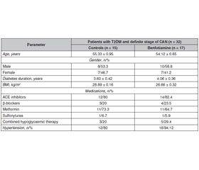
Background. Cardiac autonomic neuropathy is a serious complication of diabetes mellitus that is strongly associated with approximately five-fold increased risk of cardiovascular mortality. Cardiac autonomic neuropathy manifests itself in a spectrum of things, ranging from resting tachycardia and fixed heart rate to the development of silent myocardial infarction. The significance of diabetic cardiac autonomic neuropathy has not been fully appreciated and there is no unified treatment algorithm. The purpose was to investigate the effects of benfotiamine on the heart rate variability, the corrected QT interval, QT dispersion and spatial QRS-T angle in patients with type 2 diabetes mellitus and cardiac autonomic neuropathy. Materials and methods. Thirty-two patients with type 2 diabetes mellitus and definite stage of cardiac autonomic neuropathy were allocated into two treatment groups: control (n = 15) received traditional antihyperglycaemic therapy; group 2 (n = 17) — benfotiamine 300 mg/day for three months in addition to standard treatment. Results. It was found that benfotiamine contributed to an increase in the sum of the squares of differences between adjacent normal-to-normal intervals, pNN50 (Δ% = +45.90 ± 7.91 %, p < 0.05), high-frequency component of heart rate variability during the active (Δ% = +25.80 ± 5.58 %, p < 0.05) and passive periods of the day (Δ% = +21.10 ± 4.17 %, p < 0.05), led to a decrease in the corrected QT interval (Δ% = –7.30 ± 1.36 %, p < 0.01), QT dispersion (Δ% = –27.7 ± 9.0 %, p < 0.01) and spatial QRS-T angle (Δ% = –24.4 ± 10.2 %, p < 0.01). Conclusions. The positive influence of benfotiamine suggests the feasibility of its administration to patients with type 2 diabetes mellitus and definite stage of cardiac autonomic neuropathy.


Ключевые слова

цукровий діабет 2-го типу; автономна ней­ропатія серця; бенфотіамін; варіабельність ритму серця; коригований інтервал QT; просторовий кут QRS-T

сахарный диабет 2-го типа; автономная нейропатия сердца; бенфотиамин; вариабельность ритма сердца; корригированный интервал QT; пространственный угол QRS-T

type 2 diabetes mellitus; cardiac autonomic neuropathy; benfotiamine; heart rate variability; corrected QT interval; spatial QRS-T angle

Introduction

Cardiac autonomic neuropathy (CAN) is a serious complication of diabetes mellitus (DM) that is strongly associated with approximately five-fold increased risk of cardiovascular mortality. CAN manifests itself in a spectrum of things, ranging from resting tachycardia and fixed heart rate (HR) to the development of silent myocardial infarction [1–3]. The development of CAN is associated with the lesion of the autonomic nervous system (ANS) and may be accompanied by coronary ischemia, arrhythmias, silent myocardial infarction, severe orthostatic hypotension and sudden death syndrome [1–4]. Based on the CAN Subcommittee of the Toronto Consensus Panel on Diabetic Neuropathy [1] CAN is defined as the impairment of cardiovascular autonomic control among patients with established DM following the exclusion of other causes. Cardiac autonomic neuropathy among type 2 DM (T2DM) patients that is characterized by lesion of nerve fibers in the sympathetic and parasympathetic divisions of the ANS is diagnosed unsatisfactorily [5–9]. The reduced heart rate variability (HRV) observed in patients with diabetic CAN might indicate lower parasympathetic activity since time-domain measures of HRV and most frequency-domain indices of HRV are an expression of vagal activity [3, 10].
Hyperinsulinaemia can induce reversible prolongation of QTc (corrected QT for HR) in healthy subjects, hyperglycaemia and acute hypoglycaemia can induce the prolongation of QTc in both healthy and diabetic patients [11]. In patients with DM, prolongation of QTc was found during nocturnal hypoglycaemia that supports an arrhythmic basis for the dead in bed syndrome [12].
The spatial QRS-T angle, defined as the angle between the mean QRS and T vectors, indicates the main orientation of electrical heart activity during ventricular depolarization and repolarization, and has recently become an area of research interest. A wider QRS-T angle reflects an abnormal arrangement of ventricular repolarization and has been considered as a strong and independent risk factor for cardiac morbidity and mortality compared to other traditional cardiovascular risk factors and electrocardiographic (ECG) risk indicators such as the length of the QT interval [13–15]. The spatial QRS-T angle has recently been shown to be a strong and independent predictor of cardiac mortality in various groups of patients such as those with coronary artery disease (CAD), heart failure, T2DM, and elderly subjects [9, 15–17].
Therefore, the problem of effective treatment of CAN is particularly relevant. Pathogenetic treatment of CAN includes: balanced diet and physical activity; reducing insulin resistance; optimization of glycaemic control; treatment of dyslipoproteinaemia; correction of metabolic abnormalities in myocardium; prevention and treatment of thrombosis; use of aldose reductase inhibitors, gamma-linolenic acid, acetyl-L-carnitine, antioxidants, omega-3 polyunsaturated fatty acids, vasodilators, fat-soluble vitamin B1 (benfotiamine (BFT)), aminoguanidine; symptomatic treatment of concomitant diseases and syndromes (hypertension, coronary heart disease, heart failure, arrhythmias) and others [3, 18–21].
The purpose was to evaluate the effects of benfotiamine, a lipid-soluble thiamine derivative with higher bioavailability than that of thiamine, on the heart rate variability and the corrected QT interval, QT dispersion and spatial QRS-T angle in patients with type 2 diabetes mellitus and definite stage of cardiac autonomic neuropathy.

Materials and methods

To explore the effectiveness of some above-mentioned compounds, we examined 32 patients with T2DM and definite stage of CAN aged 50–59 years with disease duration of 1–6 years and median glycated haemoglobin A1c (HbA1c) 7.10 ± 0.40 %. Clinical characteristics of patients with T2DM and definite stage of CAN are given in Table 1.
CAN was diagnosed according to previously proposed criteria [1, 3].
The work was done according to the principles of the Declaration of Helsinki II and was approved by the medical ethics committee of Danylo Halytsky Lviv National Medical University (Minutes No. 1 dated August 29, 2016). All participants signed an informed consent prior to their inclusion in the study.
Patients with T2DM and definite stage of CAN were allocated into two treatment groups: group 1 (n = 15, controls) received traditional antihyperglycaemic therapy; group 2 (n = 17) — BFT 300 mg/day in addition to standard treatment. The duration of the therapy was 3 months.
The concentration of glucose in the blood was determined by the glucose oxidase method while HbA1c level was assessed using a highly sensitive method of ion exchange liquid chromatography with D-10 analyzer and Bio-Rad reagents (United States).
Resting 12-lead surface ECG with a paper speed of 25 mm/s and a signal size of 10 mm/mV was recorded in the morning. We performed resting ECG analysis, which included measurement of the following parameters: heart rhythm, HR, conduction intervals, and Holter-ЕCG (ЕС-3Н recorder, Labtech, Hungary) analysis included measurement of 24-h ECG, circadian indexes and following HRV parameters [22, 23]: standard deviation of all normal-to-normal (NN) intervals (SDNN), standard deviation of the means of all NN intervals for all 5-min segments of the entire recording (SDANNi), the square root of the mean of the sum of the squares of differences between adjacent NN intervals (RMSSD), percentage of adjacent NN intervals that differ from each other by more than 50 ms (pNN50, %), the high-frequency component of HRV (HF), the low-frequency component of HRV (LF), the very low-frequency component of HRV (VLF), ratio of low to high frequency power components (sympathetic/parasympathetic ratio (LF/HF)). QTc was calculated by dividing the QT interval by the square root of the preceding NN interval time series (Bazett’s formula: QTc = QT/√NN) [24]. QT dispersion (QTd) was calculated as the difference between the maximum and minimum QTc. QRS-T angle is an ECG-derived measure of the difference in mean vectors of depolarization and repolarization. The absolute difference between the frontal QRS wave axis and T-wave axis was defined as frontal planar QRS-T angle. If such a difference exceeded 180°, the difference was calculated by subtracting from 180° [25].
Statistical analysis was based on the variation method using statistical parametric t-test, nonparametric Wilcoxon test, and Fisher’s, Pearson’s correlation coefficients. Data are presented as mean ± standard error of the mean (SEM). All tests were performed using the ANOVA (MicroCal Origin v. 8.0) software. Statistical significance was set at p < 0.05.

Results

We found that in patients with T2DM and definite stage of CAN, treatment hadn’t statistically significant influence on HbA1c level (p > 0.05).
The features of the time-domain HRV parameters in patients with T2DM and definite stage of CAN after BFT administration are given in Table 2.
Obtained results could prove that treatment with BFT in patients with T2DM and definite stage of CAN led to a significant decrease in the pNN50 and did not affect the SDNN, SDANNi and RMSSD (Table 2).
Changes in the spectral heart rate variability parameters during the active period in patients with T2DM and CAN after 3 months of BFT therapy are given in Table 3.
Obtained results of this study could prove that treatment with BFT is accompanied by a significant increase in HF parameters during the active period compared to patients in control group (Table 3).
Сhanges in the spectral HRV parameters during the passive period in patients with T2DM and definite stage of CAN after 3 months of BFT therapy are given in Table 4.
It was found out that treatment with BFT is accompanied by a significant increase in HF parameters during the passive period compared to patients in control group (Table 4).
The features of the QTc, QTd and spatial QRS-T angle in patients with T2DM and advanced stage of CAN after treatment with BFT are given in Table 5.
Obtained results of this study could prove that prescription of BFT is accompanied by a more significant decrease in QTc, QTd and QRS-T angle compared to patients in control group (Table 5).
As a result of our studies, it was found that treatment with BFT contributed to a decrease in resting tachycardia (110 to 96 beats/min; p < 0.05), improvement of subjective feeling and an increase in tolerance to exercise loading. In addition, the majority of the patients with diabetic polyneuropathy (DPN) had a decrease and/or disappearance of pain, paresthesia, reduced frequency of muscle cramps, improvement and/or restoration of tactile, vibration and temperature sensitivity.

Discussion

Dysautonomia is a broad term that describes any disease or malfunction of the ANS. Evidence has been produced to indicate that various forms of mild to moderate vitamin deficiencies result in functional changes in the ANS. It is hypothesized that the predictable loss of efficiency in oxidative metabolism is the key to understanding the association of dysautonomia with many different diseases. Mild to moderate hypoxia and/or thiamine deficiency (TD) both give rise to exaggeration of centrally controlled mechanisms involved in all survival reflexes, mediated normally through a balanced reaction of the ANS and endocrine system. Together with dietary excesses, sugar consumption, particularly, appears to be responsible for triggering long-term disease related to the synthesis and use of cellular energy. Failure of the ANS cholinergic neurotransmission might follow from TD and/or other cofactors involved in glucose metabolism, exposing the organism to adrenal medullary release of epinephrine. Chronic exposure to moderate and severe hypoxia increases the activity of the ANS and adrenal medulla and TD induces an early functionally significant central muscarinic cholinergic lesion in rat studies [26].
Diabetes mellitus might be considered a thiamine-deficient state, if not in absolute terms then at least relative to the increased requirements deriving from accelerated and amplified glucose metabolism in non-insulin dependent tissues that, like the vessel wall, are prone to complications [27, 28]. The conventional indicator of thiamine sufficiency, erythrocyte transketolase (TKT) activity, is masked in clinical diabetes by increased levels of thiamine transporter-1 and thiamine transporter-2 proteins. The deficiency of thiamine in clinical DM may increase vascular fragility to the adverse effects of hyperglycaemia and, thereby, the risk of developing microvascular complications. Inhibition of TKT activity, and subsequent downregulation of the hexose monophosphate shunt, resulting in the accumulation of glyceraldehyde 3-phosphate, fructose 6-phosphate, and dihydroxyacetone phosphate, may be at least one mechanism in the development of diabetes-induced vascular damage and other comorbidities [29–31].
Thiamine and its derivatives have been demonstrated to prevent the activation of the biochemical pathways (increased flux through the polyol pathway, formation of advanced glycation end products (AGEs) pathways, activation of protein kinase C (PKC), and increased flux through the hexosamine biosynthesis pathway induced by hyperglycaemia in DM). Thiamine definitively plays an important role in the diabetic endothelial vascular diseases (micro- and macroangiopathy), lipid disorders, retinopathy, nephropathy, cardiomyopathy, and neuropathy [32, 33]. Thiamine acts as a coenzyme for TKT and for the pyruvate dehydrogenase and alpha-ketoglutarate dehydrogenase complexes, enzymes which play a fundamental role in intracellular glucose metabolism. TKT and glucose-6-phosphate dehydrogenase, the rate-limiting enzymes of the pentose phosphate pathway, are inhibited in the diabetic heart under basal conditions [34].
Experiences from cardiology indicate that long-term increases in HRV and reduction in sudden cardiac death have only been shown with lipophilic agents that readily penetrate the blood nerve/blood brain barrier. In accordance with these observations, experimental data indicate a preventive effect of BFT on the development of CAN [35, 36]. High-dose therapy of thiamine and BFT inhibited AGEs accumulation in the peripheral and reversed diabetic neuropathy potentially by reducing the levels of triose phosphates via activation of TKT [28, 37].
The evidence about positive thiamine effects was confined to hydrophobic thiamine metabolites that fulfill an important function under oxidative stress (OS) and nitrosyl stress. Thiamine protects nervous tissue probably by inhibiting nitric oxide (NO)-dependent tyrosine nitration and subsequent formation of dityrosine and interprotein tyrosine-tyrosine crosslink [31, 38]. Cardiac OS is involved in heart failure that is induced by thiamine deprivation in rats. These findings suggest that thiamine modulates OS [33]. Nitric oxide synthase is an enzyme that is involved in the synthesis of NO, which regulates a variety of important physiological responses, including cell migration, the immune response, and apoptosis. Endothelial nitric oxide synthase and NO may play an important role in attenuating cardiac remodeling and apoptosis. Benfotiamine reduces OS and activates endothelial nitric oxide synthase to enhance the generation and bioavailability of NO, and it subsequently improves the integrity of vascular endothelium to prevent sodium arsenite-induced experimental vascular endothelial dysfunction [33, 38].
Thiamine supplementation can prevent hyperglycaemia-induced reduction in cell replication and proliferation as well as decreasing AGEs formation. BFT has been shown to prevent an increase in markers of hexosamine biosynthesis pathway activity, intracellular AGEs formation, intracellular PKC activity and the nuclear factor kappa B activation seen with in vitro hyperglycaemic damage [36]. Oral BFT in combination with the antioxidant alpha-lipoic acid treatment normalizes production of angiopoietin-2, a marker of increased intracellular methylglyoxal in endothelial cells, which contribute to AGEs formation, and N-acetylglucose-modified protein, a marker of hexosamine biosynthesis pathway activity [36]. Treatment with BFT has been shown to reduce activation of the polyol pathway of glucose metabolism and to increase TKT expression in the presence of hyperglycaemia [36]. Activation of AGE receptors in DM, found on cardiomyocytes, pericytes, and podocytes, stimulates post-receptor signaling, intracellular reactive oxygen species formation, and altered gene expression, leading to vascular damage [39].
Benfotiamine supplementation (100 mg/kg/day) for 14 weeks in streptozotocin-induced diabetic mice completely corrected hyperglycaemia-induced disruptions in Ca2+ homeostasis and mechanical functioning of cardiomyocytes [39].
In our previous investigations we have found that in patients with T2DM and definite stage of CAN, QRS-T (78.30 ± 1.95°; p < 0.001), QTc (431.40 ± 2.94 ms; p < 0.001) and QTd (53.70 ± 1.49 ms; p < 0.01) were prolonged compared to patients without CAN [9]. An association between CAN and QT interval prolongation was demonstrated in many studies and it may predispose to sudden death in DM. Increased QTd was also suggested as a marker of diabetic autonomic neuropathy. Most of the data regarding QT interval and diabetic CAN are obtained in type 1 DM, with only few studies in T2DM [40].
The pathogenesis of QTc prolongation is multifactorial and includes imbalance in cardiac sympathetic innervation, intrinsic metabolic and electrolytic myocardial changes, left ventricular hypertrophy, CAD, and genetic factors could lead to QTc prolongation [41]. The day-night modulation of the QT/relative risk relation — on 24-h ECG recordings — was altered in CAN patients free of CAD, left ventricular dysfunction, or hypertrophy, with a reversed day-night pattern and an increased nocturnal QT rate dependence [34]. Reversible QTc prolongation may be induced by hyperinsulinaemia in healthy subjects, by hyperglycaemia and acute hypoglycaemia in both healthy and diabetic subjects [11, 41, 42]. In type 1 DM patients, prolonged QTc was shown to occur frequently during nocturnal hypoglycaemia and was associated with cardiac rate/rhythm disturbances. These findings support an arrhythmic basis for the dead in bed syndrome and possibly a provocative role of hypoglycaemia-induced sympathetic activation in cardiovascular events [1, 4, 12].
P. Valensi et al. demonstrated that changes in QTc can be considered as markers of cardiovascular autonomic dysfunction and as an important component in the potential prognostic value of the risk of arrhythmias [42]. Preserving the function of the parasympathetic nervous system in T2DM patients with CAN has a protective function, and the predominance of the sympathetic nervous system or the imbalance of LF/HF is harmful to the electrophysiological activity of the myocardium and may lead to changes in QRS-T [13, 14].
The spatial QRS-T angle was independently associated with glycaemic control, dyslipidaemia, and left ventricular myocardial performance in the diabetic subjects [13]. In the Rotterdam study, spatial QRS-T angle values ≥ 105° found in 20 % of patients with T2DM were associated with increased risk of cardiovascular mortality and sudden cardiac death. A spatial QRS-T angle < 75° was also significantly associated with increased risk for all clinical outcomes [43].
One recent study demonstrated that the spatial QRS-T angle is significantly wider in subjects with T2DM and CAN [14]. Moreover, presence and severity of CAN were the strongest predictors of the spatial QRS-T angle values. HRV parameters were significantly and independently associated with the spatial QRS-T angle, and explained almost 50 % of its variability, suggesting the presence of a common pathophysiological ground linking the structural, functional and electrical myocardial disturbances in DM.
The results of our study showed that the prescription of BFT in the treatment of patients with T2DM and definite stage of CAN for 3 months contributed to a decrease in the QTc, QTd and QRS-T angle. Therefore, BFT may have therapeutic potential for neurological diseases by inhibiting inflammatory mediators and enhancing anti-inflammatory factor production [21, 33, 44].
The identification of the association of polymorphisms related to the genes of thiamine and TKT with DPN might be a first step in determining a DPN genetic risk profile with potential therapeutic repercussions. There is a mode–rate evidence from preclinical experimental models that high-dose thiamine and BFT: 1) inhibit hexose monophosphate, AGEs formation, and diacylglycerol-PKC through the TKT activation; 2) target various surrogate markers of hyperglycaemia-induced pathological processes; 3) can delay the progression of microangiopathic complications [45, 46].

Conclusions

1. The administration of benfotiamine for 3 months promotes an increase in parasympathetic link activity, particularly in pNN50, HF parameters during the active and passive periods of the day, and a decrease in the QTc, QTd, QRS-T angle. Obtained results suggest the feasibility of its use in the comprehensive treatment of patients with T2DM and definite stage of CAN.
2. The mechanism of BFT influence on diabetic CAN pathogenesis is not well-known. Thus, further investigations aimed at understanding the mechanism of action and confirming the beneficial effect of BFT on biochemical parameters, dynamics of independent cardiovascular tests, daily monitoring of electrocardiography, arterial wall stiffness parameters among patients with T2DM and definite stage of CAN are needed to validate this clinical findings.
Conflict of interests. There are no ethical/legal conflicts involved in the article. There are no conflicts of interest, including specific financial interests and relationships and affiliations relevant to the subject of this manuscript.
Participation of authors: V.A. Serhiyenko — collection of material, statistical data analysis, writing the manuscript text; V.B. Segin — management of the research, development of the study design, editing of the manuscript text; L.M. Serhiyenko — management of the research, development of the study design; A.A. Serhiyenko — management of the research, editing of the manuscript text. All authors contributed equally to the review. All authors have read and approved the final version of the manuscript.
Funding. No grant or funds have been received for this study.

Список литературы

  1. Spallone V., Ziegler D., Freeman R., Bernardi L., Frontoni S., Pop-Busui R., Stevens M. et al. Cardiovascular autonomic neuropathy in diabetes: clinical impact, assessment, diagnosis, and management. Diabetes Metab. Res. Rev. 2011. 27(7). 639-53. doi: 10.1002/dmrr.1239.
  2. Serhiyenko V.A., Serhiyenko A.A. Cardiac autonomic neuropathy: risk factors, diagnosis and treatment. World J. Diabetes. 2018. 9(1). 1-24. doi: 10.4239/wjd.v9.i1.1.
  3. Spallone V. Update on the impact, diagnosis and management of cardiovascular autonomic neuropathy in diabetes: what is defined, what is new, and what is unmet. Diabetes Metab. J. 2019. 43(1). 3-30. doi: 10.4093/dmj.2018.0259.
  4. Serhiyenko V.A., Serhiyenko A.A. Diabetic cardiac autonomic neuropathy. In: Saldaña J.R., ed. Diabetes Textbook: Clinical Principles, Patient Management and Public Health Issues. Basel: Springer, Cham. Springer Nature Switzerland AG. 2019, Section 53. 825-850. Online ISBN 978-3-030-11815-0. doi: 10.1007/978-3-030-11815-0.
  5. Vinik A.I., Erbas T., Casellini C.N. Diabetic cardiac autonomic neuropathy, inflammation and cardiovascular disease. J. Diabetes Investig. 2013. 4(1). 4-18. doi: 10.1111/jdi.12042.
  6. Vinik A.I., Nevoret M.L., Casellini C., Parson H. Diabetic neuropathy. Endocrinol. Metab. Clin. North Am. 2013. 42(4). 747-87. doi: 10.1016/j.ecl.2013.06.001.
  7. Dimitropoulos A., Tahrani A.A., Stevens M.J. Cardiac autonomic neuropathy in patients with diabetes mellitus. World J. Diabetes. 2014. 1(5). 17-39. doi: 10.4239/wjd.v5.i1.17.
  8. Pop-Busui R., Boulton A.J.M., Feldman E.L., Bril V., Freeman R., Malik R.A., Sosenko J.M. et al. Diabetic neuropathy: a position statement by the American Diabetes Association. Diabetes Care. 2017. 40(1). 136-54. doi: 10.2337/dc16-2042.
  9. Serhiyenko V., Serhiyenko A. Diabetic cardiovascular neuropathy. Stavropol: Logos Publishers, 2018. doi: 10.18411/dia012018.49.
  10. Bernardi L., Spallone V., Stevens M., Hilsted J., Frontoni S., Pop-Busui R., Ziegler D. et al. Toronto Consensus Panel on Diabetic Neuropathy. Methods of investigation for cardiac autonomic dysfunction in human research studies. Diabetes Metab. Res. Rev. 2011. 27(7). 654-64. doi: 10.1002/dmrr.1224.
  11. Prince C.T., Secrest A.M., Mackey R.H., Arena V.C., Kingsley L.A., Orchard T.J. Cardiovascular autonomic neuropathy, HDL cholesterol, and smoking correlate with arterial stiffness markers determined 18 years later in type 1 diabetes. Diabetes Care. 2010. 33(3). 652-57.
  12. Desouza C.V., Bolli G.B., Fonseca V. Hypoglycemia, diabetes, and cardiovascular events. Diabetes Care. 2010. 33(6). 1389-94. doi: 10.2337/dc09-2082.
  13. Voulgari C., Moyssakis I., Perrea D., Kyriaki D., Katsilambros N., Tentolouris N. The association between the spatial QRS-T angle with cardiac autonomic neuropathy in subjects with type 2 diabetes mellitus. Diabet. Med. 2010. 27(12). 1420-9. doi: 10.1111/j.1464-5491.2010.03120.x.
  14. Voulgari C., Pagoni S., Tesfaye S., Tentolouris N. The spatial QRS-T angle: implications in clinical practice. Curr. Cardiol. Rev. 2013. 9(3). 197-210. doi: 10.2174/1573403X113099990031.
  15. Gungor M., Celik M., Yalcinkaya E., Polat A.T., Yuksel U.C., Yildirim E. et al. The value of frontal planar QRS-T angle in patients without angiographically apparent atherosclerosis. Med. Princ. Pract. 2017. 26(2). 125-31. doi: 10.1159/000453267.
  16. Walsh J.A. 3rd, Soliman E.Z., Ilkhanoff L., Ning H., Liu K., Nazarian S., Lloyd-Jones D.M. Prognostic value of frontal QRS-T angle in patients without clinical evidence of cardiovascular disease (from the Multi-Ethnic Study of Atherosclerosis). Am. J. Cardiol. 2013. 112(12). 1880-4. doi: 10.1016/j.amjcard.2013.08.017.
  17. Raposeiras-Roubin S., Virgos-Lamela A., Bouzas-Cruz N., López-López A., Castiñeira-Busto M., Fernández-Garda R. et al. Usefulness of the QRS-T angle to improve long-term risk stratification of patients with acute myocardial infarction and depressed left ventricular ejection fraction. Am. J. Cardiol. 2014. 113(8). 1312-19. doi: 10.1016/j.amjcard.2014.01.406.
  18. Deli G., Bosnyak E., Pusch G., Komoly S., Feher G. Diabetic neuropathies: diagnosis and management. Neuroendocrinology. 2013. 98(45). 267-80. doi: 10.1159/000358728.
  19. Serhiyenko V.A., Serhiyenko A.A. Diabetic cardiac autonomic neuropathy: Do we have any treatment perspectives? World J. Diabetes. 2015. 6(2). 245-58. doi: 10.4239/wjd.v6.i2.245.
  20. Serhiyenko V.A., Serhiyenko A.A. Omega-3 polyunsaturated fatty acids in the treatment of diabetic cardiovascular autonomic neuropathy: a review. In: Moore S.J., ed. Omega-3: dietary sources, biochemistry and impact on human health. New York: Nova Science Publishers, 2017. 79-154. ISBN: 978-1-53611-824-7 print. Online ISBN: 978-1-53611-839-1.
  21. Serhiyenko V., Hotsko M., Snitynska O., Serhiyenko A. Benfotiamine and type 2 diabetes mellitus. MOJ Public Health. 2018. 7(1). 00200. doi: 10.15406/mojph.2018.07.00200.
  22. Heart rate variability. Standards of measurement, physiological interpretation, and clinical use. Task Force of the European Society of Cardiology and the North American Society of Pacing and Electrophysiology. Circulation. 1996. 93(5). 1043-65. PMID: 8598068.
  23. Adamec J., Adamec R. ECG Holter: guide to electrocardiographic interpretation. New York: Springer-Verlag US, 2008. doi: 10.1007/978-0-387-78187-71.
  24. Bazett H.C. An analysis of the time-relations of electrocardiograms. Ann. Noninvasive Electrocardiol. 1997. 2(2). 177-94. doi: 10.1111/j.1542-474X.1997.tb00325.x.
  25. Borleffs C.J., Scherptong R.W., Man S.C., van Welsenes G.H., Bax J.J., van Erven L., Swenne C.A. et al. Predicting ventricular arrhythmias in patients with ischemic heart disease: clinical application of the ECG-derived QRS-T angle. Circ. Arrhyth. Electrophysiol. 2009. 2(5). 548-54. doi: 10.1161/CIRCEP.109.859108.
  26. Lonsdale D. Dysautonomia, a heuristic approach to a revised model for etiology of disease. Evid. Based Complementary Altern. Med. 2009. 6(1). 3-10. doi: 10.1093/ecam/nem064.
  27. Berrone E., Beltramo E., Solimine C., Ape A.U., Porta M. Regulation of intracellular glucose and polyol pathway by thiamine and benfotiamine in vascular cells cultured in high glucose. J. Biol. Chem. 2006. 281(14). 9307-13. doi: 10.1074/jbc.M600418200.
  28. Rašić-Milutinović Z., Gluvić Z., Peruničić-Peković Z., Miličević D., Lačković M., Penčić B. Improvement of heart rate variability with benfothiamine and alpha-lipoic acid in type 2 diabetic patients — pilot study. J. Cardiol. Ther. 2014. 2(1). 1-7. doi: 10.12970/2311-052X.2014.02.01.9.
  29. Oh S.H., Witek R.P., Bae S.H., Darwiche H., Jung Y., Pi L., Brown A., Petersen B.E. Detection of transketolase in bone marrow-derived insulin-producing cells: benfotiamine enhances insulin synthesis and glucose metabolism. Stem Cells Dev. 2009. 18(1). 37-45. doi: 10.1089/scd.2007.0255.
  30. Stirban A., Pop A., Tschoepe D. A randomized, double-blind, crossover, placebo-controlled trial of 6 weeks benfotiamine treatment on postprandial vascular function and variables of autonomic nerve function in type 2 diabetes. Diabet. Med. 2013. 30(10). 1204-08. doi: 10.1111/dme.12240.
  31. Pácal L., Kuricová K., Kaňková K. Evidence for altered thiamine metabolism in diabetes: is there a potential to oppose gluco- and lipotoxicity by rational supplementation? World J. Diabetes. 2014. 5(3). 288-95. doi: 10.4239/wjd.v5.i3.288.
  32. González-Ortiz M., Martínez-Abundis E., Robles-Cervantes J.A., Ramírez-Ramírez V., Ramos-Zavala M.G. Effect of thiamine administration on metabolic profile, cytokines and inflammatory markers in drug-naïve patients with type 2 diabetes. Eur. J. Nutr. 2011. 50(2). 145-9. doi: 10.1007/s00394-010-0123-x.
  33. Luong K.V., Nguyen L.T. The impact of thiamine treatment in the diabetes mellitus. J. Clin. Med. Res. 2012. 4(3). 153-60. doi: 10.4021/jocmr890w.
  34. Katare R.G., Caporali A., Emanueli C., Madeddu P. Benfotiamine improves functional recovery of the infarcted heart via activation of pro-survival G6PD/Akt signaling pathway and modulation of neurohormonal response. J. Mol. Cell. Cardiol. 2010. 49(4). 625-38. doi: 10.1016/j.yjmcc.2010.05.014.
  35. Kempler P. Review: autonomic neuropathy: a marker of cardiovascular risk. Br. J. Diabetes Vasc. Dis. 2003. 3(2). 84-90. doi: 10.1177/14746514030030020201.
  36. Boghdadi M.A., Afify H.E., Sabri N., Makbout K., Elmazar M. Comparative study of vitamin B complex combined with alpha lipoic acid versus vitamin B complex in the treatment of diabetic polyneuropathy in type 2 diabetic patients. Clin. Exp. Pharmacol. 2017. 7(4). 241.
  37. Stracke H., Gaus W., Achenbach U., Federlin K., Bretzel R.G. Benfotiamine in diabetic polyneuropathy (BENDIP): results of a randomised, double blind, placebo-controlled clinical study. Exp. Clin. Endocrinol. Diabetes. 2008. 116(10). 600-5. doi: 10.1055/s-2008-1065351.
  38. Pacal L., Tomandl J., Svojanovsky J., Krusova D., Stepankova S., Rehorova J. et al. Role of thiamine status and genetic variability in transketolase and other pentose phosphate cycle enzymes in the progression of diabetic nephropathy. Nephrol. Dial. Transplant. 2011. 26(4). 1229-36. doi: 10.1093/ndt/gfq550.
  39. Moss C.J., Mathews S.T. Thiamin status and supplementation in the management of diabetes mellitus and its vascular comorbidities. Vitam. Min. 2013. 2. 111. doi: 10.4172/vms.1000111.
  40. Pillai J.N., Madhavan S. Cardiac autonomic neuropathy and QTc Interval in type 2 diabetes. Heart India. 2015. 3(1). 8-11. doi: 10.4103/2321-449X.153279.
  41. Veglio M., Borra M., Stevens L.K., Fuller J.H., Perin P.C. The relation between QTc interval prolongation and diabetic complications: The EURODIAB IDDM Complications Study Group. Diabetologia. 1999. 42(1). 68-75. doi: 10.1007/s001250051115.
  42. Valensi P., Johnson N.B., Maison-Blanche P., Extramania F., Motte G., Coumel P. Influence of cardiac autonomic neuropathy on heart rate dependence of ventricular repolarization in diabetic patients. Diabetes Care. 2002. 25(5). 918-23. doi: 10.2337/diacare.25.5.918.
  43. Kardys I., Kors J.A., Van der Meer I., Hofman A., Van der Kuip D.A., Witteman J.C. Spatial QRS-T angle predicts cardiac death in a general population. Eur. Heart J. 2003. 24(14). 1357-64. doi: 10.1016/s0195-668x(03)00203-3.
  44. Shoeb M., Ramana K.V. Anti-inflammatory effects of benfotiamine are mediated through the regulation of the arachidonic acid pathway in macrophages. Free Radic. Biol. Med. 2012. 52(1). 182-90. doi: 10.1016/j.freeradbiomed.2011.10.444.
  45. Koufopoulos G., Pafili K., Papanas N. Correlation between type 2 diabetes mellitus and ankle-brachial index in a geographically specific Greek population without peripheral arterial disease. Mìžnarodnij endokrinologìčnij žurnal. 2019. 15(7). 9-14. doi: 10.22141/2224-0721.15.7.2019.186053.
  46. Ziegler D., Schleicher E., Strom A., Knebel B., Fleming T., Nawroth P., Haring H.U. et al. Association of transketolase polymorphisms with measures of polyneuropathy in patients with recently diagnosed diabetes. Diabetes Met. Res. Rev. 2016. 33(4). e2811. doi: 10.1002/dmrr.2811.

Вернуться к номеру