
Международный эндокринологический журнал Том 15, №7, 2019
Вернуться к номеру
Активатори транскетолази як новий підхід до лікування: їх значення в патогенезі та лікуванні діабетичних мікросудинних ускладнень
Авторы: Singh K., Yuzvenko T., Kogut D.
Ukrainian Research and Practical Centre for Endocrine Surgery, Transplantation of Endocrine Organs and Tissues of the Ministry of Health of Ukraine, Kyiv, Ukraine
Рубрики: Эндокринология
Разделы: Справочник специалиста
Версия для печати
В оглядовій статті наведено аналіз літературних даних про вплив тіаміну і його похідних на метаболізм у клітинах і його роль у патогенезі ускладнень цукрового діабету. Розглянуто особливості перетворення неактивної форми, яка потрапляє в організм людини з продуктами харчування та препаратами, в активну форму тіаміну. Також розглянуті особливості метаболічних шляхів тіаміну і його похідних. Описані особливості алітіаміну і прикладне значення його метаболізму в клітинах для лікування ускладнень цукрового діабету, зокрема діабетичної полінейропатії. Дані, наведені в цій статті, допоможуть клініцистам зрозуміти важливість та необхідність використання в своїй практиці препаратів, що містять тіамін.
В обзорной статье приведен анализ литературных данных о влиянии тиамина и его производных на метаболизм в клетках и его роли в патогенезе осложнений сахарного диабета. Рассмотрены особенности преобразования неактивной формы, которая попадает в организм человека с продуктами питания и препаратами, в активную форму тиамина. Также рассмотрены особенности метаболических путей тиамина и его производных. Описаны особенности аллитиамина и прикладное значение его метаболизма в клетках для лечения осложнений сахарного диабета, в частности диабетической полинейропатии. Данные, приведенные в этой статье, помогут клиницистам понять значимость и необходимость использования в своей практике препаратов, содержащих тиамин.
The review article presents an analysis of the literature on the effect of thiamine and its derivatives on the metabolism in cells and its role in the pathogenesis of complications of diabetes mellitus. The features of the conversion of an inactive form that enters the human body with food and drugs into the active form of thiamine are considered. The metabolic pathways of thiamine and its derivatives are also clarified. The article describes the features of allithiamine and the practical importance of its metabolism in cells for the treatment of complications of diabetes mellitus, in particular, diabetic polyneuropathy. The data in this article will help clinicians understand the relevance and necessity of using drugs containing thiamine in their practice.
цукровий діабет; мікросудинні ускладнення; транскетолаза; діабетична полінейропатія; тіамін; алітіамін; огляд
сахарный диабет; микрососудистые осложнения; транскетолаза; диабетическая полинейропатия; тиамин; аллитиамин; обзор
diabetes mellitus; microvascular complications; transketolase; diabetic polyneuropathy; thiamine; allithiamines; review
Introduction
Approximately 425 million people in the world have diabetes mellitus (DM), out of which around 1.3 million cases of DM were registered in Ukraine. The global pre–valence of DM in individuals over the age of 18 years has risen from 4.7 % in 1980 to 8.5 % in 2014 [1]. According to the WHO, diabetes was the seventh leading cause of death in 2016.
DM is characterized by hyperglycemia-induced tissue damage as established in the Diabetes Control and Complications Trial and the U.K. Prospective Diabetes Study. Generally, the damaging effects of hyperglycemia are differentiated into microvascular complications (diabetic retinopathy, neuropathy and nephropathy) and macrovascular complications (coronary artery disease, peripheral artery disease and stroke). The effects of hyperglycemia are not seen in all cells of the body, but are distinct only in certain types of cells: neurons and Schwann cells in peripheral nerves, capillary endothelial cells, mesangial cells in the renal glomerulus due to their inability to effectively maintain a constant level of glucose, in contrast most cells are able to reduce the transport of glucose when exposed to hyperglycemia [2, 3].
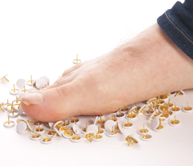
Diabetic sensory polyneuropathy (DSPN) is amongst the most common long-term complications of diabetes [7]. It has been estimated that 50 % of diabetic patients will have neuropathy within 25 years of their disease history [8]. The increased risk of non-traumatic foot amputations and lower extremity disease in diabetic patients has been attributed to DSPN [9, 10]. An early diagnosis helps identify patients at risk before the onset of disabling complications. Despite its common occurrence, DSPN shows considerable variability in prevalence. This can be attributed to the differences in definitions of neuropathy and the tests used for the evaluation [11].
Information on existing mechanisms
The exact mechanism of damage to the peripheral nerves due to hyperglycemia is not understood fully but is likely to be related to sorbitol accumulation, advanced glycation end products (AGE) and oxidative stress [5]. Peripheral neuropathy may manifest in various forms: sensory, focal, multifocal, autonomic. Although neurological signs and symptoms are recommended diagnostic tools [12], subjectivity, lack of reproducibility and proficiency of the examiner leads to inaccuracies [13]. Moreover, asymptomatic neuropathy is common, pre–sent in up to 50 % of cases, and clinical features do not always correlate with the severity of pathological changes [14]. Other forms of neuropathy may replicate the picture in diabetic sensory neuropathy and mononeuropathy [5]. Chronic inflammatory polyneuropathy, vitamin B12 deficiency, hypothyroidism, and uremia should be considered while evaluating diabetic polyneuropathy in patients [15].
Diabetic autonomous neuropathy is also one of the important factors leading to morbidity and mortality in diabetic patients. Neuropathic changes may manifest as gastroparesis, constipation, erectile dysfunction, bladder dysfunction, resting tachycardia, silent ischemia, and even cardiac arrest [15].
The pathobiology of diabetic complications as a unifying mechanism has been explained in the Banting lecture in 2004. Hyperglycemia-induced mitochondrial superoxide production activates the four damaging pathways by inhibiting GAPDH that leads to microvascular diabetic complications.
The polyol pathway activation resulted from the increased glucose flux causes sorbitol accumulation in cells. Increased sorbitol is considered as the underlying mechanism in the development of diabetic microvascular complications [4, 5].
Advanced glycation end products pathway leads to the increase of the major intracellular AGE precursor me–thylglyoxal [4]. These substances have been linked with the formation of microaneurysms and pericyte loss [5].
Protein kinase C pathway is activated due to the inhibition of GAPDH activity that leads to the increase in glyceraldehyde-3-phosphate which is a precursor to diacylglycerol [4].
Hexosamine pathway realizes by the conversion of fructose-6-phosphate by the enzyme GFAT to UDP-N-acetylglucosamine [4], which results in increased expression of TGF β1 and plasminogen activator inhibitor-1 [6].
Prophylaxis and treatment
According to the generally approved guidelines and practices, the primary goal of the therapy in DM patients is to control symptoms and prevent worsening of microvascular diabetic complications. Although some stu–dies suggest that tight glycemic control and avoidance of glycemic excursions may improve symptoms in diabetic peripheral neuropathy [5].
One of the novel approaches for the prevention and treatment of diabetic complications based upon the unifying mechanism for the pathogenesis of diabetic complications is using the transketolase activators [4]. Fructose-6-phosphate and glyceraldehyde-3-phosphate are also final products of transketolase reaction in the pentose phosphate shunt [16]. As glycolytic intermediates can flow to the pentose phosphate pathway depending on the concentration of substrate presented to transketolase and as in the case of diabetic patients with hyperglycemia, activation of transketolase diverts the flux of glycolytic intermediates away from the damaging pathways [5]. The enzyme transketolase requires a co-factor thiamine for activation.
Thiamine diphosphate, also known as thiamine pyrophosphate or cocarboxylase is a water-soluble vitamin. Thiamine diphosphate is the coenzyme for five key me–tabolic enzymes: mitochondrial pyruvate dehydrogenase complexes, oxoglutarate dehydrogenase complexes, branched-chain 2-oxo acid dehydrogenase complexes, 2-hydroxyacyl-CoA lyase 1 as well as the cytosolic trans–ketolase.
Allithiamine (benfotiamine) is a member of the class of lipophilic thiamine derivatives. The unique properties of the allithiamines result from the opening of the thiazole ring of thiamine upon reaction with sulphur compounds. Upon dephosphorylation of benfotiamine in the intestinal tract [17, 18] a lipophilic molecule is produced, which readily diffuses across cell membranes and is absorbed much better than water-soluble thiamine salts. This property allows for greater absorption both in the intestines and in target tissues as compared with thiamine itself. Following penetrating into the cell, the molecule undergoes catalytic reduction by intracellular sulfhydryl compounds and/or enzymatic debenzoylation [17], closing the thiazole ring and releasing the active thiamine into the cell and circulation [17].
In one study, thiamine activated transketolase by 25 % in arterial endothelial cells, benfotiamine activated transketolase by 250 % [5].
The properties of benfotiamine and its role in cell metabolism have been described in various fundamental research papers, to name a few: Hammes H.P. et al. described that benfotiamine prevents the increase in UDP-N-acetylglucosamine, and normalizes protein kinase C activity and prevents nuclear factor kappa B activation in retina of diabetic patients [20]. Berrone E., Beltramo E. et al. demonstrated that benfotiamine corrects imbalances in the polyol pathway by decreasing aldose reductase activity, sorbitol concentrations and intracellular glucose, thereby protecting endothelial cells from glucose-induced damage [21]; benfotiamine corrects glucose-induced endothelial cell damage by normalizing cell replication rates and decreasing apoptosis [21].
The significance of benfotiamine in clinical practice has also been demonstrated in many clinical trials with variable trial designs, patient groups and history of diabetes, e.g. the BEDIP study and BENDIP study.
Conclusions
As allithiamines activate transketolase more significantly, further investigation is needed to verify the effect of allithiamines in comparison to thiamine diphosphate in terms of their affinity to THTR-1 receptor and THTR-2 receptor, which are encoded by the reduced folate carrier family.
In the Ukrainian Research and Practical Centre for Endocrine Surgery, Transplantation of Endocrine Organs and Tissues, we have also been studying the role of benfotiamine. A fundamental study regarding the efficacy of benfotiamine in treating patients with diabetic polyneuropathy in its various stages of manifestation and its effect on the expression of genes of the reduced folate carrier is being carried out.
The detailed description of the study is out of the scope of this article and will be presented in consequent articles.
Conflicts of interests. Authors declare the absence of any conflicts of interests and their own financial interest that might be construed to influence the results or interpretation of their manuscript.