
Международный неврологический журнал №7 (101), 2018
Вернуться к номеру
Case analysis of crossed cerebellar hemispheric diaschisis in acute stroke patients
Авторы: S.M. Vinychuk(1), O.Ye. Fartushna(2)
(1) — Oleksandrivska Clinical Hospital, Kyiv, Ukraine
(2) — Ukrainian Military Medical Academy, Kyiv, Ukraine
Рубрики: Неврология
Разделы: Клинические исследования
Версия для печати
Актуальність. Інсульт є другою за частотою причиною смертності у світі, поступаючись лише смертності від серцево-судинних захворювань. Концепція діашизу Монакова описує нейрофізіологічні зміни, що відбуваються на відстані від осередкового ураження головного мозку і віді-грають значну роль у вираженості гострого неврологічного дефіциту в пацієнтів з інсультом. Проте на цей час опубліковано недостатньо прoспективних клінічних досліджень, у яких проаналізовано характеристики перехресного мозочково-півкульного діашизу в пацієнтів iз гострим інсультом. Мета дослідження: визначення особливостей клінічних проявів перехресного мозочково-півкульного діашизу в пацієнтів iз гострим ішемічним інсультом й підвищення ефективності його діагностики шляхом зіставлення даних клініко-неврологічного дослідження з результатами магнітно-резонансної томографії. Матеріали та методи. Ми провели проспективне госпітальне когортне дослідження 124 пацієнтів iз гострим ішемічним інсультом, які надійшли до відділення цереброваскулярних захворювань Олександрівської клінічної лікарні Києва протягом перших 24 годин iз моменту розвитку інсульту. Усі пацієнти пройшли комплексне клініко-неврологічне, лабораторне, ультразвукове та нейровізуалізаційне обстеження. Результати. Серед 124 обстежених iз гострим ішемічним інсультом перехресний мозочково-півкульний діашиз був діагностований у 6 (4,8 %) пацієнтів. Ми проаналізували патофізіологічні, анатомічні та клінічні особливості перехресного мозочково-півкульного діашизу в пацієнтів iз гострим ішемічним інсультом. Висновки. Основним механізмом виникнення перехресного мозочково-півкульного діашизу є переривання еферентних імпульсів у мозочково-зубчастому або зубчасто-таламокортикальному шляху. Отже, ізольовані мозочкові інфаркти проявлялися не тільки атактичними розладами, але й моторними і сенсорними порушеннями, викликаними пошкодженням лобно-тім’яної кори (діашиз) протилежної півкулі головного мозку.
Актуальность. Инсульт является второй по частоте причиной смертности в мире, уступая лишь смертности от сердечно-сосудистых заболеваний. Концепция диашиза Монакова описывает нейрофизиологические изменения, которые происходят вдали от очагового поражения головного мозга и играют значительную роль в выраженности острого неврологического дефицита у пациентов с инсультом. Тем не менее в настоящее время опубликовано недостаточно прoспективных клинических когортных исследований, в которых проанализированы характеристики перекрестного мозжечково-полушарного диашиза у пациентов с острым инсультом. Цель исследования: определение особенностей клинических проявлений перекрестного мозжечково-полушарного диашиза у пациентов с острым ишемическим инсультом и повышение эффективности его диагностики путем сопоставления данных клинико-неврологического обследования с результатами магнитно-резонансной томографии. Материалы и методы. Мы провели проспективное госпитальное когортное исследование 124 пациентов с острым ишемическим инсультом, поступивших в отделение цереброваскулярных заболеваний Александровской клинической больницы Киева в течение первых 24 часов с момента развития инсульта. Все пациенты прошли комплексное клинико-неврологическое, лабораторное, ультразвуковое и нейровизуализационное обследование. Результаты. Среди 124 обследованных с острым ишемическим инсультом перекрестный мозжечково-полушарный диашиз был диагностирован у 6 (4,8 %) пациентов. Мы проанализировали патофизиологические, анатомические и клинические особенности перекрестного мозжечково-полушарного диашиза у пациентов с острым ишемическим инсультом. Выводы. Основным механизмом возникновения перекрестного мозжечково-полушарного диашиза является прерывание эфферентных импульсов в мозжечково-зубчатом или зубчато-таламокортикальном пути. Следовательно, изолированные мозжечковые инфаркты проявлялись не только атактическими расстройствами, но и моторными и сенсорными нарушениями, вызванными повреждением лобно-теменной коры (диашиз) противоположного полушария головного мозга.
Background. Stroke is the second-leading global cause of death behind the heart disease, accounting for 11.8 % of total deaths worldwide. The Monakow concept of diaschisis describes neurophysiological changes that occur distant to a focal brain lesion. Diaschisis plays a significant role in the severity of acute neurological deficit and spontaneous stroke recovery. However, currently there are not enough published prospective hospital-based cohort studies that report and analyze clinical characteristics of crossed cerebellar hemispheric diaschisis in acute stroke patients. The purpose of this study is to determine the features of the clinical manifestations of crossed cerebellar hemispheric diaschisis after acute cerebral stroke and to improve the efficiency of its diagnosis by comparing the obtained data with the results of the magnetic resonance imaging. Materials and methods. We prospectively recruited 124 acute stroke patients, who were admitted to a single department at an academic tertiary care hospital in Kyiv, Ukraine. The primary outcome was the combined incidence of stroke and diaschisis. In the secondary analyses, we evaluated pathophysiological, anatomical, and clinical features specific to crossed cerebellar hemispheric diaschisis in a cohort of acute stroke patients. Results. Among 124 selected acute stroke patients admitted to the department, 42 (33.9 %) persons were diagnosed with different forms of diaschisis. Crossed cerebellar hemispheric diaschisis was diagnosed in 6 patients. We described clinical manifestations and analyzed pathophysiological and clinical features of crossed cerebellar hemispheric diaschisis in acute stroke patients. The main mechanism of crossed cerebellar hemispheric diaschisis is an interruption of efferent impulses at the cerebellar dentate or dentate-thalamo-cortical pathway. Conclusions. Consequently, isolated cerebellar infarctions were manifested not only in ataxic disorders, but also in motor and sensory impairments due to the damage of the frontal-parietal cortex (diaschisis) of the opposite hemisphere of the brain.
перехресний мозочково-півкульний діашиз; інсульт мозочка; мозочок; дистантний діашиз; форми діашизу; клінічні прояви; діагностика; клінічний випадок
перекрестный мозжечково-полушарный диашиз; мозжечковый инсульт; мозжечок; дистантный диашиз; формы диашиза; клинические проявления; диагностика; клинический случай
crossed cerebellar hemispheric diaschisis; cerebellar stroke; cerebellum; distant diaschisis; forms of diaschisis; clinical manifestations; diagnosis, case report
Introduction
Materials and methods
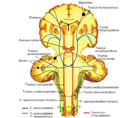
Results and discussion
/18.jpg)
/18_2.jpg)
Conclusions
1. Benjamin E.J. On behalf of the American Heart Association Council on Epidemiology and Prevention Statistics Committee and Stroke Statistics Subcommittee. Heart disease and stroke statistics 2018 update: a report from the American Heart Association / E.J. Benjamin, S.S. Virani, C.W. Callaway [et al.] // Circulation. — 2018. — Vol. 137(12). — P. e67-e492.
2. Fartushna O.Ye. Brain injury in patients with acute TIA: clinical features in different TIA subtypes / O.Ye. Fartushna, S.M. Vinychuk // Международный неврологический журнал. — 2017. — № 3(89). — С. 13-18.
3. Feigin V.L. Global burden of stroke / V. L. Feigin, B. Nor-rving, G.A. Mensah // Circulation research. — 2017. — № 120(3). — Р. 439-448.
4. Lees R. Vascular cognitive impairment/vascular dementia. The pattern of cognitive impairment in stroke survivors with carotid stenosis / R. Lees, F. McGrane, O. Fartushna, N.M. Broomfield, T.J. Quinn, K. Dani, K. Forbes, J. Dawson // International Journal of Stroke. — 2014. — № 9. — P. 323-324.
5. Wilkins E. European cardiovascular disease statistics 2017 / Е. Wilkins, L. Wilson, K. Wickramasinghe [et al.]. — Brussels: European Heart Network, 2017. — 188 p.
6. Евтушенко С.К. Новые факторы риска развития инсульта у лиц молодого возраста / С.К. Евтушенко, Д.А. Филимонов, И.С. Евтушенко // Журнал неврологии и психиатрии им. С.С. Корсакова. Спецвыпуск. — 2015. — Т. 115, № 12. — С. 3-12.
7. Віничук С.М. Рання реабілітація після гострих ішемічних порушень мозкового кровообігу / С.М. Віничук, О.Є. Фартушна // Міжнародний неврологічний журнал. — 2016. — № 8(86). — С. 34-39.
8. Фартушна О.Є. Виявлення та усунення васкулярних чинників ризику — важливий напрямок первинної профілактики транзиторних ішемічних атак та/чи інсульту / О.Є. Фартушна, С.М. Віничук // Український медичний часопис. — 2015. — № 1(105). — С. 23-27.
9. Фартушна О.Є. Актуальність проблеми цереброваскулярних захворювань, транзиторних ішемічних атак та вдосконалення їх діагностики в системі охорони здоров’я в Україні / О.Є. Фартушна, М.М. Прокопів // Проблеми військової охорони праці: Зб. наук. праць Української військово-медичної академії / За ред. проф. Білого В.Я. — К.: УВМА, 2007. — Вип. 19. — С. 335-342.
10. GBD 2016 Causes of Death Collaborators. Global, regional, and national age-sex specific mortality for 264 causes of death, 1980–2016: a systematic analysis for the Global Burden of Disease Study 2016 // The Lancet. — 2017. — № 390(10100). — Р. 1151-1210.
11. Wang H. Global, regional, and national life expectancy, all-cause mortality, and cause-specific mortality for 249 causes of death, 1980–2015: a systematic analysis for the Global Burden of Disease Study 2015 / H. Wang, M. Naghavi, C. Allen [et al.] // The Lancet. — 2016. — № 388(10053). — P. 1459-1544.
12. World Stroke Organization. Facts and Figures about Stroke. — Режим доступу: http://www.world-stroke.org/component/content/article/16-forpatients/84-facts-and-figures-about-stroke.
13. Фартушна О.Є. Транзиторні ішемічні атаки / О.Є. Фартушна, С.М. Віничук. — К.: ВД «Авіцена», 2014. — 216 с.
14. Фартушна О.Є. Епідеміологія транзиторних ішемічних атак в структурі гострих порушень мозкового кровообігу в Украї-ні та інших країнах / О.Є. Фартушна, С.М. Віничук // Міжнародний неврологічний журнал. — 2017. — № 5(91). — С. 105-111.
15. Finger S. The von Monakow concept of diaschisis: origins and perspectives / S. Finger, P.J. Koehler, C. Jagella // Archiv Neurologie. — 2004. — Vol. 61. — P. 283-288.
16. Seitz R.J. The role of diaschisis in stroke recovery / R.J. Seitz, N.P. Azari, U. Knorr [et al.] // Stroke. — 1999. — № 30(9). — P. 1844-50.
17. Vinychuk S.M. Diaschisis: brief historical review / S.M. Vinychuk, O.Ye. Fartushna // Міжнародний неврологічний журнал. — 2018. — № 4(98). — C. 11-15.
18. Виничук С.М. Диашиз при мозговом инсульте. — К.: ОЖИВА, 2017. — 64 c.
19. Віничук С.М. Історія Київської неврологічної школи / С.М. Віничук, О.Є. Фартушна. — К.: Едванс-Прінт, 2015. — 55 с.
20. Виничук С.М. Диашиз и его роль в развитии рефлекторно-двигательных расстройств при мозговом инсульте // Український медичний часопис. — 2013. — № 2. — С. 143-147.
21. Vinychuk S.M. Cerebrospinal and commissural diaschisis in acute stroke patients: case analysis / S.M. Vinychuk, O.Ye. Fartushna // Международный неврологический журнал. — 2018. — № 5(99). — C. 28-33.
22. Vinychuk S.M. Crossed cerebellar diaschisis in acute stroke patients: case analysis and report / S.M. Vinychuk, O.Ye. Fartushna // Международный неврологический журнал. — 2018. — № 6(100). — C. 15-20.
23. Kernan W.N. Guidelines for the prevention of stroke in patients with stroke and transient ischemic attack: a guideline for healthcare professionals from the American Heart Association/American Stroke Association / W.N. Kernan, B. Ovbiagele, H.R. Black [et al.] // Stroke. — 2014. — № 45. — Р. 2160-2236.
24. Aho K. Cerebrovascular disease in the community: results of a WHO collaborative study / K. Aho, P. Harmsen, S. Hatano [et al.] // Bull. World Health Organ. — 1980. — № 58. — P. 113-130.
25. Adams H.P. Classification of subtype of acute ischemic stroke. Definitions for use in a multicenter clinical trial. TOAST. Trial of Org 10172 in Acute Stroke Treatment / H.P. Adams, B.H. Bendixen, L.J. Kappelle [et al.] // Stroke. — 1993. — № 24. — P. 35-41.
26. Ringleb P. European Stroke Organisation 2008 guidelines for managing acute cerebral infarction or transient ischemic attack. Part 1 / P. Ringleb, P.D. Schellinger, W. Hacke [et al.] // Der Nervenarzt. — 2008. — № 79. — P. 936-957.
27. Sacco R.L. Guidelines for prevention of stroke in patients with ischemic stroke or transient ischemic attack: a statement for healthcare professionals from the American Heart Association/American Stroke Association Council on Stroke: co-sponsored by the Council on Cardiovascular Radiology and Intervention: the American Academy of Neurology affirms the value of this guideline / R.L. Sacco, R. Adams, G. Albers [et al.] // Stroke. — 2006. — № 37. — P. 577-617.
28. Fartushna O.Y. Emergency therapeutic approach as a secondary prevention of an acute ischemic stroke in patients with TIA / Fartushna O.Y. // XX World Neurological Congress, 12–17.11.2011. — Marrakesh, Morocco, 2011. — P. 167.
29. Fartushnaya E.E. Reducing the risk of recurrent ischemic stroke, after transient ischaemic attack along with neuroprotective and antiaggregant therapy / E.E. Fartushnaya, S.M. Vinichuk // XIV International Congress of Rehabilitation Medicine and Immunorehabilitation, 16–21.10.2009: Abstract. — Tel-Aviv, Israel, 2009. — Р. 67.
30. Віничук С.М. Диференційоване лікування транзиторних ішемічних атак — ефективний спосіб профілактики повторних гострих церебральних подій / С.М. Віничук, О.Є. Фартушна // Міжнародний неврологічний журнал. — 2014. — № 6. — С. 87-92.
31. Віничук С.М. Аторвастатин та його роль у профілактиці та лікуванні ішемічних порушень мозкового кровообігу / С.М. Віничук, О.Є. Фартушна // Здоров’я України. — 2015. — № 9. — С. 3.
32. Фартушна О.Є. Використання оптимальної дози препарату Торвакард — важливий напрямок зниження ризику розвитку повторних транзиторних ішемічних атак та/чи інсульту / О.Є. Фартушна, С.М. Віничук // Семейная медицина. — 2015. — № 3. — С. 223-227.
33. Віничук С.М. Освітні програми профілактики транзиторних ішемічних атак та/чи інсульту / С.М. Віничук, О.Є. Фартушна // Укр. мед. часопис. — 2014. — № 5. — С. 49-51.
34. Фартушна О.Є. Модифікація поведінкових чинників ризику як складова первинної профілактики транзиторних ішемічних атак та/чи інсульту / О.Є. Фартушна, С.М. Віничук // Український медичний часопис. — 2014. — № 6(104). — XІ/XІІ. — С. 42-44.
35. Фартушна О.Є. Патогенетичні підтипи транзиторних ішемічних атак: особливості неврологічної клініки, гемодинаміки та лікування [Текст]: Дис... канд. мед. наук: 14.01.15 / Фартушна Олена Євгенівна; Нац. мед. ун-т ім. О.О. Богомольця. — К., 2012. — 217 арк.: рис., табл. — Бібліогр.: арк. 187-217.
36. Переверзев И.В. Метаболические нарушения в головном мозге больных, перенесших инсульт мозжечка / И.В. Переверзев // Врач. Ежемесячный научно-практический публицистический журнал. — 2010. — № 5. — С. 77-79.
37. Crisomoto R.A. Detection of diffusion — weighted attack and minor stroke patients using acute magnetic resonance imaging / R.A. Crisomoto, M.M. Corcia, D.C. Tong // Ann. Neurol. — 2003. — Vol. 57(6). — P. 848-859.
38. Grips E. Supratentorial age-related white matter changes predict outcome in cerebellar stroke / E. Grips, O. Sedlaczek, H. Bäzner et al. // J. Cereb. Blood Flow Metab. — 2005. — Vol. 36(9). — P. 1988-1998.
39. Holmes G. The cerebellum of man / G. Holmes // Brain. — 1930. — Vol. 62. — P. 1-30.