
Журнал «Почки» Том 7, №2, 2018
Вернуться к номеру
Використання генетичного тестування при стероїд-резистентному нефротичному синдромі
Авторы: B.S. Lipska-Ziętkiewicz
Clinical Genetics Unit, Department of Biology and Genetics, Medical University of Gdańsk, Gdańsk, Poland
Рубрики: Нефрология
Разделы: Справочник специалиста
Версия для печати
Нефротичний синдром, одне з найпоширеніших захворювань нирок, що уражають пацієнтів різного віку, відрізняється характерним генетичним складом, особливо в осіб, які не відповідають на лікування глюкокортикоїдами. У результаті недавніх розробок у галузі молекулярної біології генетичне тестування стало діагностичним інструментом першої лінії при диференціальній діагностицi захворювання. У цьому міні-огляді наведені сучасні й майбутні напрямки генетичного тестування при нефротичному синдромі.
Нефротический синдром, одно из самых распространенных заболеваний почек, поражающих пациентов всех возрастов, отличается характерным генетическим составом, особенно у лиц, которые не отвечают на лечение глюкокортикоидами. В результате недавних разработок в области молекулярной биологии генетическое тестирование стало диагностическим инструментом первой линии при дифференциальной диагностике заболевания. В этом мини-обзоре представлены современные и будущие направления генетического тестирования при нефротическом синдроме.
Nephrotic syndrome, one of the most frequent kidney conditions affecting patients of all ages, has significant genetic composition, especially in individuals who do not respond to glucocorticoid treatment. The recent developments in molecular biology transformed genetic testing into the first-line diagnostic tool in differential diagnosis of the condition. In this mini review current and future directions of genetic testing in nephrotic syndrome are presented.
стероїд-резистентний нефротичний синдром; генетичне тестування; подоцитопатія
стероид-резистентный нефротический синдром; генетическое тестирование; подоцитопатия
steroid-resistant nephrotic syndrome; genetic testing; podocytopathies
Molecular basis of the nephrotic syndrome
After decades of primarily morphological diagnostic algorithms in the clinical management of the nephrotic syndrome (NS), in the last ten years we are observing the emergence of genetic testing as an important player in reaching the final diagnosis. This echoes the general trend in modern medicine, usually referred to as “personalized medicine” where treatment is tailored for each patient according to the precise characterization of the individual both inherited and environmental particularities of the disease.
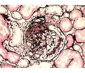
Nephrotic syndrome is one of the most frequent kidney conditions to affect children and adults. Conditions presenting clinically as NS may be either highly kidney specific, associated with extra-renal developmental malformations or occur as complication of a systemic disease [2]. The hallmark of nephrotic syndrome is malfunction of the glomerular filtration barrier (GFB). The GFB comprises three layers: podocytes, glomerular basement membrane (GBM) and the fenestrated endothelium that together make up the ultra-filter of plasma by the kidney. Recent developments in the pathogenesis of NS point toward the central role of podocytes in maintaining barrier integrity what resulted in coining the term podocytopathy (podocytopathies) when referring to the heterogeneous group of clinical entities manifesting as NS. These include both idiopathic but also clearly genetic conditions, with a well defined pattern of extra-renal manifestations.
The recommended first choice treatment of NS is glucocorticoid therapy allowing not only for disease regression in majority of cases but also for classification of patients into responders to immunosuppression that usually will have a good prognosis and non-responders, who generally progress to renal failure but have low rate of disease relapse after successful kidney transplantation. The latter group is the one with highest chances of detecting the underlying genetic defect. It should be underlined however, that about 8–10 % of genetic cases respond, at least initially, to intensified immunosuppression. This is true with respect to calcineurin inhibitors in particular. Conversely, children with multidrug-resistant sporadic disease show better renal survival than those with genetic disease [11].
The genes associated with hereditary nephrotic syndrome
As for today, there are more than 50 genes associated with NS (for the major ones see Table 1). The encoded proteins are essential for glomerular filtration barrier integrity and function, playing a role in maintaining the proper structural characteristics of the slit diaphragm, cytoskeleton, cell-matrix interactions (GBM) or regulating podocyte functioning through various signal transduction pathways and control of the energy homeostasis.
/9-1.jpg)
The communication between slit diaphragm and the podocyte cytoskeleton inloves a number of proteins of which the most important is nephrin (encoded by the NPHS1 gene), the essential component of the GFB interacting with the transmembrane protein podocin (NPHS2) and phospholipase C epsilon 1 (PLCE1). Mutations in the NPHS1 account for ~60–80 % of congenital nephrotic syndrome followed by PLCE1, WT1 and LAMB2 mutations. NPHS2 is the single most commonly mutated gene in steroid-resistant nephrotic syndrome (SRNS) of any, except for infants, age [12]. A gain of function mutations in another slit diaphragm protein, a calcium channel localized at podocyte foot process encoded by TRPC6 gene, leads to increased calcium influx and subsequent dysregulation of the actin cytoskeleton.
Podocyte cytoskeleton is responsible for maintaining their specialized shape, and this is achieved through interplay of a number of actin network proteins. Among these, the most commonly mutated in SRSN are structu–ral components encoded by ACTN4 and MYO1E and actin regulatory element encoded by INF2. Mounting evidence for the role of major GBM proteins encoded by COL4A5, COL4A3, COL4A4 genes that classically cause Alport syndrome in late-onset (adulthood) SRNS supports their inclusion in NGS-based diagnostic gene panels [6].
Genes encoding nuclear regulatory factors better known as transcription factors and alterations to nuc–lear pore complex proteins comprise another subgroup of SRNS-related genomic loci. Among these, the most frequently mutated are WT1, SMARCAL1 and LMX1B. Mutations in these genes lead to a number of developmental genetic syndromes including Denys — Drash and Frasier syndromes (WT1), Schimke immuno-osseous dysplasia (SMARCAL1) and nail-patella syndrome (LMX1B).
Finally, a group of genes encoding proteins involved in several metabolic (mitochondrial) pathways is also involved in both syndromic and idiopathic SRNS. The most common genetic entity is ADCK4-related glomerulopathy followed by other disorders affecting coenzyme Q10 biosynthesis pathway.
Prevalence of genetic forms of SRNS
So far, genetic diagnosis explains not more than 1/3 of SRNS with the highest detection rate in familial, consanguineous and young (congenital/infantile) patients that drops to < 10 % in sporadic adults ([10]; PodoNet own data — unpublished). Accordingly, the mode of inheritance and the age of onset are the most important clues suggestive for a particular gene defect. These factors however are not so easy to be applied clinically. The mode of inheritance can be clearly defined in a small subset of cases only, as the majority of patients present with negative family history or with just one affected relative. More to the point, due to non-full penetrance and variable expressivity, some family members carrying pathogenic variants in late-onset autosomal dominant genes might easily be overlooked. The second of the postulated factors pointing towards hereditary nature of the condition is the age of onset. Indeed, the younger the patient is, the higher the chances to detect causative mutations, with the highest detection rates of 60–80 % in congenital NS. Nonetheless, mutations in the same gene can be responsible for SRNS from infancy to adulthood as it is the case with the most frequently affected NPHS2 gene. In the past, a few age-wise mutational screening algorithms have been proposed, however two large NGS studies ([10]; PodoNet — own data) only partially reproduced such age-related patterns. Therefore, the current recommendation is to test all SRNS-related genes at once (using for instance NGS exome sequencing or large-gene NGS panel approach). For countries, where local medical health insurance policies obstruct such an expensive testing, two step cost-effective approaches based on the mutational yield might be proposed. Here, prior to NGS, patients could be screened at low cost for mutations in the entire coding sequence of NPHS2 and exons 8,9 of WT1 as these represent roughly ~50 % of genetic diagnoses.
Genotype-phenotype correlations
From clinical perspective, the genotype-phenotype correlations in SRNS are not so straightforward and easy to interpret clinically. In the past, only patients presenting a full-blown phenotype (for instance a classic Denys — Drash syndrome: an individual with sex-reversal, bilate–ral Wilms tumor and infantile SRNS with DMS lesions on biopsy) were catching the attention of the “gene” hunters and could have the genetic diagnosis established. That resulted in a rather stringent diagnostic criteria and limited recommendations for genetic testing. Today, in the era of next generation sequencing offering robust parallel sequencing of numerous genes, more and more oligosymptomatic patients reach diagnosis without need of waiting for the syndrome to fully develop. That has reversed the management strategy — today genotype anticipates phenotype. Clinical geneticists play the role of a wise fairy, able to predict yet clinically silent manifestations, potential involvement of other organs and systems, as well as foretell treatment strategy and the eventual outcome. The above mentioned Denys — Drash syndrome can be used as an illustration of the aforesaid: an infant presenting NS and diagnosed with a WT1 pathogenic mutation not only will NOT have steroid treatment initiated nor kidney biopsy performed, but rather will be put on a short-track towards kidney transplantation. Moreover, such child will also undergo an intensive oncological surveillance program aimed at Wilms tumor and gonadoblastoma detection and will be subject to in-depth endocrine assessment aimed at detection of a disorder of sex differentiation along with karyotype testing [8]. As another, even more successful example, ADCK4-related glomerulopathy can serve. It is an important novel genetic entity of insidious onset at adolescence with mild to moderate proteinuria and absence of relevant edema in the majority of patients resulting in an already advanced chronic kidney disease at the time of diagnosis in half of the cases. ADCK4 gene mutations are more common in Asian (for instance Turkish, Chinese) populations. Despite belonging to coenzyme Q10 (CoQ10) biosynthesis pathway disorders, a subgroup of mitochondropathies, in ADCK4-related disease extra-renal manifestations are scarce and, if present, relatively mild. CoQ10 glomerulopathy is the first hereditary form of SRNS for which a causative molecular therapy is potentially available. Oral CoQ10 supplementation may reverse proteinuria and stabilize kidney function if applied early in the di–sease course [5]. At PodoNet Consortium, through fa–mily tes–ting we were able to identify ten yet-asymptomatic siblings eligible for CoQ10 supplementation who showed significant decrease in proteinuria with no effect on estimated glomerular filtration rate while on treatment [1].
There is, however, another side to the phenotype-prediction performance in SRNS. As shown for instance in the case of SMARCAL1 mutations, a significant fraction of patients carrying pathogenic variants in this gene does not manifest clinically the phenotype consistent with Schimke immuno-osseous dysplasia. In the recently published series of 34 patients with SMARCAL1 mutations detected through PodoNet Registry, extrarenal symptoms and patient survival varied widely and correlated with the type of mutation, whereas the proteinuric glomerulopathy invariably progressed to end-stage kidney disease within the first two decades of life [9]. Other examples include certain pathogenic variants in WT1 (further discussed in [8]), the mutations affecting p.R246 codon of LMX1B [3] or some mutations in exons 2-4(6) of INF2 [4]. It can be expected that the detection rate of milder variants will increase with routine NGS gene panel screening hampering genetic counseling. It is also observed that patients harboring the same mutation may present completely different disease dynamics. Such a phenomenon, most likely attributed to yet unidentified factors, either modifying genes, epigenetic alterations or environmental factors, was for example observed in a series of patients — compound heterozygotes carrying p.R229Q non-neutral polymorphism and c.1032delT mutation in NPHS2 gene [7]. Among 11 patients (5 fa–milies), all coming from a closed ethnical group occupying a relatively isolated geographical region of Pomerania in Northern Poland, a significant inter- and intra-family clinical heterogeneity was observed with the age of onset ranging from infancy till late adolescence.
Where do we go from now
It is generally acknowledged that roughly half of the SRNS is due to an underlying genetic defect. 2/3 of these will have the underlying monogenic disorder identified. The remaining cases most likely represent effect of podocyte injury by an unknown circulating factor, cytokine imbalance or other immune processes. These, although considered immunological conditions, may still have some genetic background which could play part in their pathogenesis. In brief, it cannot be excluded that some rare genomic variants determine susceptibility or resistance to immune stimuli as well as the character of the subsequent response. Actually, it is now believed that monogenic inheritance of abnormalities in podocyte-specific genes disrupting filter function is only part of the story. In fact, in some patients pathological changes are observed focally (not in all glomeruli) and/or segmentally (only in parts of the glomerulus), what suggests that there was an environmental stressor, for instance a viral infection, that exercised initial localized insult in a genetically predisposed individual [2]. Such multifactorial pattern of inheritance, otherwise called “moderately complex trait”, provides explanation why the observed genotype-phenotype correlations in NS are far from being consistent. It is eagerly anticipated that the robust analyses of NGS and clinical data from large, well characterized NS patient cohorts will allow better understanding of the observed considerable genetic heterogeneity. These analy–zes have to take into account not only the gene-sequencing information which is already used in clinical (diagnostic) setting but also a number of factors under research, such as epigenetic modifications, imprinting, miRNAs, etc. Only then we will be able to have a full picture of the NS pathogenesis and hence be able to predict precisely the phenotype and tailor adequate treatment modalities.
Note from the author
Diagnostic genetic testing of the entire coding sequence of NPHS2 and exons 8,9 of WT1 that account for ~50 % of hereditary SRNS can be performed in the Clinical Genetics Laboratory at the Medical University Polyclinics, Gdańsk, Poland (Smoluchowskiego 17, 80214 Gdańsk, Poland). Material: 2 × 2.5 ml (3 ml) or 1 × 5 ml EDTA blood sample shipped overnight with some ice-coolers (NO dry ice required). Further details: b.lipska@gumed.edu.pl.
Acknowledgments
The research leading to these results has received funding from the European Community’s Seventh Framework Programme (FP7/2007-2013) under grant agreement No. 2012-305608 (EURenOmics), from Bundesministerium für Bildung und Forschung (BMBF) through the e-Rare initiative (PodoNet) and from the Polish Ministry of Science and Education (grant N402631840).
Conflicts of interests. Authors declare the absence of any conflicts of interests that might be construed to influence the results or interpretation of their manuscript.