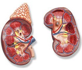
Primary chronic adrenal insufficiency (CAI, Addison’s disease) is a pathological condition associated with a decrease in the production of glucocorticoids and mineralocorticoids by the cortical layer of the adrenal glands [1]. The main cause of the disease is autoimmune adrenalitis (about 90 % of patients), other etiologic factors include infectious, iatrogenic and genetic [2, 3]. The main clinical manifestations of the disease include electrolyte disorders (hyponatremia and hyperkalemia, hypoglycemia, hyperpigmentation of the skin and mucous membranes) [2, 3]. Quite often in the endocrino–logy clinic, states associated with the combination of several endocrine, autoimmune pathogenesis-related mechanisms, as well as non-endocrine autoimmune diseases, are occured. The autoimmune polyglyandular syndrome (APS) of type 1, including mucous-cutaneous candidiasis, hypoparathyroidism and CIA, is described by D. Waiteker [4]. Often in this condition, there may be pernicious anemia, alopecia, vitiligo, bronchial asthma, glomerulonephritis, chronic autoimmune hepatitis.
It is believed that the disease develops gradually with the consequent emergence of hypoparathyroidism, CAI, later it can manifest primary hypogonadism. It is advi–sable to perform a diagnostic search after detecting one autoimmune disease of possible other syndrome components. APS type 2 was described by M. Schmidt in 1926, it included chronic autoimmune thyroiditis (AIT) and CAI [5]. Another variant of the APS type 2 is Carpenter’s syndrome (a combination of CAI, type 1 diabetes mellitus (DM) and AIT, described in 1964 by C. Carpenter) [6]. In 1981 N. Neufeld defined APS type 2 as a combination of CNN, DM type 1 and/or AIT [7].
A clinical case of observation of a patient with Carpenter’s syndrome, which had clinical features and presented certain difficulties in diagnosis, is shown. Patient P., 47 years old, hospitalized in the endocrinology department of the KI ‟RK Endocrine Dispensary” ZRC on 18th May, 2017 with complaints of pronounced general weakness, fatigue, frequent hypoglycemic conditions, a decrease in body weight of 15 kg over the past 6 months, nausea, vomiting, epigastric pain, right hypochondrium and pyloroduodenal zone.
From anamnesis
DM type 1 has been diagnosed since 1992, from the onset of the disease on insulin therapy. Periodically he noted the development of hypoglycemic conditions. According to the patient, over the past year, there has been an increase in the development of hypoglycemic conditions, twice hypoglycemic coma, which required medical intervention. At time of hospitalization he took insulin therapy in intensify regimen: Insuman Rapid in the mor–ning 2 IU, in the noon 2 IU, in the evening 2 IU, at 22.00 Insuman Basal — 6 IU. The dose of insulin has significantly decreased over the last year. Glycemia fluctuations from 2.9 to 29.2 mmol/l. From 12th April 2017 to 25th April 2017 he was in-patient in the gastroenterological department of the Zaporizhzhia Regional Clinical Hospital with diagnosis of peptic ulcer, active phase, moderate severity, chronic pyloric ulcer, with increased acid-forming function of the stomach, chronic pancreatitis. At discharge, positive dynamics were noted. In the anamnesis: arterial hypertension for 2 years (he takes enalapril 5 mg per day).
Objective examination
The general condition of mode–rate severity, he’s conscious, the skin is clean, earthy gray colour, BMI is 19.6, the location of fatty tissue is even, breathing rate — 16 per min, the percussion is clear over the lungs, breathing is vesicular, rales are absent, cardiac activi–ty is rhythmic of the heart, blood pressure 110/80 mm Hg, the abdomen is mild, moderately painful in epigastrium and in the pyloroduodenal zone palpatory. The liver and spleen are not enlarged. Stool and diuresis are normal. The pastosity of the shins and feet is marked, pulsation over the peripheral arteries of the lower limbs is reduced.
Laboratory data
Blood count (19.05.2017): hemoglobin — 124 g/l, RBC — 3,7 • 1012/l, WBC — 4,1 • 109/л, ESR — 19 mm/hr, eosinophils — 1 %, stab — 0 %, segmented — 65 %, lymphocytes — 31 %, monocytes — 3 %.
Biochemistry (19.05.2017): total cholesterol — 3.5 mM/l, triglycerides — 1.42 mM/L, VLDL choleste–rol — 0.56 mM/L, LDL cholesterol — 2.28 mM/L, urea — 5.2 mM/L, creatinine — 116,6 uM/l, GFR — 58, total bilirubin — 11.0 uM/l, direct bilirubin — 2.8 uM/l, thymol test — 0.99, AST — 0.27, ALT — 0.54, total protein — 56,1 g/l, potassium — 3,97 mM/l, sodium — 132 mM/l.
Hormonal examination: 26.05.2017: fТ4 — 15,2 mM/l (10–25), ТSH — 1,7 mIU/ml, Аb-ТPO — 10,0 (0–30) ME/ml, 25.05.2017: ACTH — 837 mM/l (7,2–63,3), cortisol — 0,88 mM/l (6,2–19,4).
Glycemic profile (08:00–11:00–16:00–20:00):
— 20.05.2017 — 4,1–9,2–11,1–5,6 mM/l;
— 23.05.2017 — 10,4–5,8–6,0–8,0 mM/l;
— 26.05.2017 — 7,4–14,0–6,2–12,7 mM/l.
Urinalysis:
19.05.2017: gravity — 1006, WBC — 0–2 in area vision, protein, ketones absent, singular flat epithelium.
22.05.2017: urinalysis by Nechiporenko: WBC — 500/mm3, RBC absent, protein absent.
24.05.2017: daily glucosuria absent, daily proteinuria absent.
27.05.2017: microalbuminuria — 89 mg/day.
Instrumental data
ECG (19.05.2017). HR — 80 beats per min, the vol–tage is lowered, sinus rhythm, electrical axis of the heart is not rejected.
X-ray of the gastrointestinal tract (29.05.2017): ulcer of duodenal bulb, chronic pancreatitis. Examination of barium passage: the whole weight of barium in the distal ileum, cecum and ascending colon.
Fiber-optic gastroduodenoscopy (24.05.2017): Atrophic gastropathy, erosive bulbitis, scar deformation of duodenal bulbs.
CT of abdominal organs (25.05.2017): CT signs of abdominal and retroperitoneal adenopathy, nodular hyperplasia of the left adrenal gland (uneven contours due to the presence in its body node spherical shape with sharp, smooth contours, relatively homogeneous structure with a diameter of 6 mm), simple cysts of left kidney.
Narrow specialist consultations
Neurologist (18.05.2012): dismetabolic encephalopathy 1, cerebroasthenic syndrome, diabetic distal symmetric polyneuropathy of the lower limbs, sensory-motor form (NSS 3, NDS 3), chronic course.
Ophthalmologist (02.06.2017): the optic nerve disc is pale pink, the borders are clear, the arteries are narrowed, and moderate angiosclerosis. Salus 1. Microane–urysms of venous vessels, microhemorrhages. In the macular area without features. Conclusion: Nonprolife–rative diabetic retinopathy of both eyes.
Gastroenterologist (22.05.2017): Peptic ulcer, inactive phase, chronic pyloric ulcer in the stage of scarring, chronic pancreatitis in the stage of unstable remission. Recommended: X-ray of the stomach with repeated examination. 25.05.2017: Peptic ulcer, active phase, chronic erosive gastroduodenitis in the acute stage, associated with H.pylori.
Angiosurgeon (06.06.2017): diabetic angiopathy of the arteries of the lower extremities.
Oncourologist (26.05.2017): hyperplasia of the left adrenal gland, oncouropathology is absent.
Comments
Picture of gastric dyspepsia (nausea and vomiting) were associated with the clinic of peptic ulcer (the history of the disease was taken into account, the diagnosis of the active phase of the disease was confirmed after additional examination and consultation of the gastroenterologist). During treatment in the hospital, there was a lack of compensation for the level of glycemia, despite the ongoing correction of insulin doses. At the same time, the decrease in the level of arterial pressure at the peak of hypoglycemia (up to 80/40 mm Hg) attracted attention. After additional examination, the development of primary insufficiency of the adrenal cortex was revealed. Mass in the left adrenal gland is most likely a hormonal non-active benign tumor (incidentaloma), or has a relationship to the systemic pathological process associated with lymphadenopathy in the abdominal cavity, which remained diagnostically unclear.
Diagnosis: Diabetes mellitus type 1, severe form labile course with a tendency to hypoglycemic states, decompensation. Diabetic nonproliferative retinopathy of both eyes. Diabetic peripheral distal symmetrical polyneuropathy (NSS 3, NDS 3), sensory-motor form, chronic course. Diabetic angiopathy of the lower extremities, ІІ degree. Diabetic nephropathy III degree. CKD III. Chronic adrenal insufficiency, moderate severity, decompensation. Dysmetabolic encephalopathy, 1 degree, cerebroasthenic syndrome. Peptic ulcer, active phase, chronic ulcer of the duodenal bulb, associated with H.pylori, with increased acid-producing function of the stomach. Arterial hypertension, I degree, 2 stage, high cardiovascular risk, hypertensive angiopathy of the retina. Nodular hyperplasia of the left adrenal gland, hormone-inactive. Intraabdominal and retroperitoneal lymphadenopathy of the unexplained genesis.
Treatment
The patient continued to receive insulin therapy with correction of insulin doses under the control of the glycemia level. Dexamethasone 4 mg IV was administered droply, then medrol in dose 16 mg at 7.00 and 11.00. In time of introduction of glucocorticoid drugs, the patient noted improvement in the condition in the form of improvement in appetite, an increase in the level of blood pressure to 120/80 mm Hg. Hypoglycemic conditions didn’t repeat. So far as patient had low blood pressure, antihypertensive therapy wasn’t applied. The patient continued the complex therapy of peptic ulcer according to the previous recommendation. Further recommendations on the therapy of chronic adrenal insufficiency (correction of the dose of the drug depending on the level of electrolytes of blood, ACTH) are given.
Features of the current Carpenter syndrome in this patient:
1. Since 1992 there was diagnosed diabetes mellitus type 1 in the patient (he has always treating with intensified insulin therapy regimen, at the time of recent examination he had BMI — 19.6 kg/m2).
2. The color of the skin is not typical for adrenal insufficiency (earthy-gray), which made it necessary to exclude renal pathology.
3. Long history of diabetes mellitus type 1 without changes from other organs.
4. As an agent contributing to the manifestation of CAI was the exacerbation of peptic ulcer.
5. For a long time, the absence of hypotension clinic against the background of hypertension syndrome.
Thus, the defined clinical case showed possible difficulties in the diagnosis of combined autoimmune endocrine pathology and internal diseases, when the clinical picture of chronic adrenal insufficiency completely masked the development of diabetes mellitus and peptic ulcer. It should be remembered the need for a diagnostic search for adrenal pathology in patients with diabetes mellitus type 1 with frequent unexplained hypoglycemic conditions and a decrease in blood pressure.
Conflicts of interests. Authors declare the absence of any conflicts of interests that might be construed to influence the results or interpretation of their manuscript.

