
Журнал «Боль. Суставы. Позвоночник» 2 (18) 2015
Вернуться к номеру
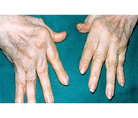
Changes in Bone Mineral Density of the Distal Forearm and Development of Erosive Joint Damage at Hand Joints MRI in Patients with Early Rheumatoid Arthritis
Авторы: Vershynina D. - Ivano-Frankivsk National Medical University, Ivano-Frankivsk, Ukraine
Рубрики: Ревматология, Травматология и ортопедия
Разделы: Медицинские форумы
Версия для печати
Статья опубликована на с. 98
Introduction. Chances for better prognosis in rheumatoid arthritis (RA) much depend on the beginning of an adequate basic treatment for early, non–erosive stage disease (no later than 3 months after the start of the articular syndrome) as a new therapeutic paradigm involves the diagnosis of RA in the most initial stages of the disease. Newest methods of X–ray diagnosis of lesions of bones and joints allow using of a number of modern techniques for early diagnosis of rheumatoid arthritis, defining the degree of destructive changes and assessing of the progression of the disease. The introduction into clinical practice of MRI significantly enhanced the diagnosis of inflammatory changes of a wrist joint and a hand. Currently, the issues of early visualizing of the changes in the joints and bone which can be used as predictors of future joints structural damage are being discussed.
Aim. To assess changes in the bone mineral density (BMD) in patients with early rheumatoid arthritis (ERA) and how the BMD is related to the erosive changes of the wrist on MRI.
Methods. Тhe study involved 112 patients with early rheumatoid arthritis (ERA) who had suffered of articular syndrome for 1 year (average 10.6 ± 2.2 months). The diagnosis was established against the RA classification criteria EULAR/ARC 2010. Mean age of the patients was 44.8 ± 7.4 years. Clinical and laboratory studies were included: rheumatologist examination, DAS28 test, ESR, concentrations of rheumatoid factor (RF) test, anti–cyclic citrullinated peptide (anti–CCP) and anti–modified citrullinated vimentin (anti–CMV) tests. Bone mineral density (BMD) of the distal radius and lumbar spine (L1–L4) were assessed by DEXA Challenger (DMS, France). MRIs of the patient's dominant wrist and 2nd — 5th metacarpophalangeal (MCP) joints were obtained using 1.5T MRI MAGNETOM Espree (Siemens) with contrast enhancement. The average score was determined for synovitis (0–9 for wrist joint, and 0–21 for wrist plus MCP joints), bone oedema (osteitis, 0–69) and bone erosions (0–230) using the RAMRIS system. Statistical analysis of the results was also undertaken.
Results. The DEXA showed bone loss in patients with ERA. When analyzing the lumbar spine osteoporosis was found in 8 patients (7.14 %) and osteopenic syndrome was identified in 33 patients (29.4 %). In the study of the distal radius the more substantial bone loss was established. Thus, osteoporosis was diagnosed in 29 patients (25.9 %), osteopenic syndrome — in 67 patients (59.8 %). The BMD decrease at the distal radius correlated with DAS28 (r = –0.67; p < 0.001), ESR (r = –0.44; p < 0.05) and number of swollen joints (r = –0.40; p < 0.01). As for BMD of the lumbar spine, only the correlation with DAS28 was found. Osteopenia and osteoporosis were more frequently identified in patients with RF (+), anti–CCP (+) and ant–CMV (+). All patients with seropositive rheumatoid arthritis showed a significant decrease in bone mineral density at both the lumbar spine and distal radius. Bone erosions were identified with standard radiography in 28 patients. Most of the erosions were located in the bones of the wrist and in 7 patients they were located in the head section of metacarpal bones. vdHS system demonstrated the score of 15.0 ± 24.6 points of average erosions. Detection of erosions on radiographs correlated with the presence of synovitis hand joints. At the same time all patients with radiographic erosions suffered of the decrease in BMD of the distal radius (r = –0.72; p < 0.001). However, assessment of the changes in mineral density of the lumbar spine did not demonstrate such correlation. MRI of the dominant wrist revealed the following MR symptoms: swelling of the bone marrow — in 102 patients (91.07 %), synovitis — in 86 patients (76.8 %), erosion — in 71 patients (63.4 %). MRI detected the erosions 2.5 times more cases than the standard radiography. Strong correlation between BMD of the distal radius and the presence of erosions detected by MRI (r = –0.72; p < 0.001), and lumbar spine BMD and erosions (r = –0.48; p < 0.01) was established.
Conclusion. The results of this study prove that the decrease in BMD of the distal radius and lumbar spine correlates with the erosions of the dominant wrist and 2nd — 5th MCP joints detected by standard radiographs and MRI. Thus, the BMD changes can be predicted early by the development of erosive process in patients with ERA. The early loss of a wrist bone tissues, measured in the first year of the disease using the DRA is an independent predictor of erosive progression.