
Журнал «Здоровье ребенка» 6 (57) 2014
Вернуться к номеру
Rosai dorfman disease in child
Авторы: Veselyy S.V., Abdullin R.F., Chercun A.V., Inozemcev I.N., Litovka V.K., Lepihov P.A., Kondratenko E.G. -
Donetsk National Medical University named after М. Gorky; Regional Children’s Clinical Hospital, Donetsk
Рубрики: Педиатрия/Неонатология
Разделы: Справочник специалиста
Версия для печати
Rosai Dorfman disease, also known as sinus histiocytosis with massive lymphadenopathy, is a benign histiocytic proliferative condition of unsubstantiated etiology that most often presents as bilateral painless cervical lymphadenopathy.
The neck lymph nodes are the most frequently involved, followed by inguinal, axillary and mediastinal lymph nodes. The most common extranodal sites are the skin, the upper respiratory tract and bones. The skin is commonly affected; half of these patients have another associated extranodal site. Orbit and ocular glove involvement have been reported, usually as a retroorbitary mass and proptosis. The characteristic histological feature is lymphophagocytosis which is specific for this syndrome.
Head and neck manifestations of Rosai Dorfman are diverse but most commonly present as massive cervical lymphadenopathy. The differential diagnosis is made with lymphoreticular malignancies such as lymphomas, Hodgkin's disease, malignant histiocytosis and monocytic leukemia, all of which have similar histopathological features. Atypias in cytology and the aggressive clinical course establish the diagnosis in most cases. Other histiocytoses, such as rhinoscleromas, Wegener's granulomatosis, may also be included in the differential diagnosis. We report the case of Rosai Dorfman disease in a 3-years old child, which underwent surgical exploration and documented the histological diagnosis of Rosai Dorfman disease.
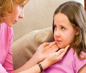
3 years boy, was hospitalized in the regional children’s clinical hospital of Donetsk. Boy complained on tumor formation on the left side of the neck. Child received conservative treatment in the community with no positive effect. Had to consult a hematologist, and then sent to the clinic.
Birth weight 3,800 gr. Been vaccinated, grows and develops by age. State of health is satisfactory. Skin clean. In the lungs, breathing puerile. Heart activity is rhythmic. Abdomen is not distended, soft, accessible to palpation in all departments. In the left side of the neck, in the upper third of the projection of the sternocleidomastoid muscle is determined lumpy rounded formation, about 4.0 cm in diameter.
In the clinic the child examined. In the blood analysis - no pathology. Chest X-ray - without pathology. Ultrasonography neck - under the sternocleidomastoid muscle visualized enlarged lymph nodes, one of which is the size of 31 × 18 × 23 mm, round, hypoechoic, homogeneous. Ultrasonography of the abdomen - focal changes spleen, diffuse liver changes.
Clinical diagnosis was exhibited. Lymphogranulomatosis?
03.04.2014 operative treatment was made. Skin incision 4.0 cm. Dissected surface fascia, lateral abdomen sternocleidomastoid muscle medially set aside. In the audit found that the tumor presented conglomerate lymph nodes 4,0 × 4,5 cm, in a capsule, whitish-gray color. Conglomeration of lymph nodes removed. Wound drained. Wound sutured.
Puncture and bone marrow aspiration from the sternum and the iliac crests, trepanobiopsy both iliac bones were done.
Histological conclusion (№ 1619-34): histology and immunophenotype cells correspond sinus histiocytosis with massive lymphadenopathy (Rosai Dorfman disease).
Myelogram: red germ reduced among lymphocyte atypical lymphocytes are found in a small number with irregular shape of the nucleus and chromatin structure furrowed.
The postoperative period was uneventful. Sutures were removed on the 8th day. The wound healed by first intention. Child was discharged for further treatment in the department of oncohematology.
Rosai Dorfman disease сmay starts from emergence of lymphadenopathy, mainly neck, with extranodal lesion areas (most often - the spleen). To clarify the diagnosis is expedient to use ultrasound, computed tomography. Definitive diagnosis is established after surgical removal (biopsy) conglomerate of lymph nodes, histological and histochemical study.
1. L'vov A.N., Voloshchuk Y.N., Varshavskyy V.A., Horbacheva Yu.V., Bobko S.Y. Synusniy hystyotsytoz (bolezn' Rozay – Dorfmana): klynycheskoe nablyudenye. // Vestnyk dermatolohyy y venerolohyy – 2011. – #5 – S.115–120.
2. Rayt D., Эddys B., Leonh Э. Morfolohycheskaya dyahnostyka patolohyy lymfatycheskykh uzlov. – M. Med. lyt., 2008. – 176s
3. Duval M, Nguyen VH, Daniel SJ. Rosai-Dorfman disease: an uncommon cause of massive cervical adenopathy in a two-year-old female. Otolaryngol Head Neck Surg. 2009 Feb;140(2):274-5. doi: 10.1016/j.otohns.2008.10.041. PubMed PMID: 19201306.
4. Lima FB, Barcelos PS, Constâncio AP, Nogueira CD, Melo-Filho AA. Rosai-Dorfman disease with spontaneous resolution: case report of a child. Rev Bras Hematol Hemoter. 2011;33(4):312-4. doi: 10.5581/1516-8484.20110083. PubMed PMID: 23049324; PubMed Central PMCID: PMC3415761.
5. Juskevicius R, Finley JL. Rosai-Dorfman disease of the parotid gland: cytologic and histopathologic findings with immunohistochemical correlation. Arch Pathol Lab Med. 2001 Oct;125(10):1348-50. PubMed PMID: 11570913.