Журнал «Здоровье ребенка» 4 (55) 2014
Вернуться к номеру
Cardiac echinococcosis in a child (a case from practice)
Авторы: G.E. Sukhareva
Рубрики: Педиатрия/Неонатология
Разделы: Справочник специалиста
Версия для печати
echinococcosis, heart, children.
Echinococcosis is considered a chronic disease due to the damage of organs and tissues of the human body caused by the immature (larval) form of tapeworm, Echinococcus. About 350 cases of echinococcus cysts in the heart have been described in the literature.
We would like to present one of such cases: a child Sasha F., aged 11. As the case-history had it, the boy had had the pain in the heart which was thought to be caused by physical exertion for 6 months before marked clinical manifestations of the disease appeared. The condition worsened precipitously when the child developed acute allergic reaction accompanied by itching, urticaria, and fever up to 39-40°C. The boy was admitted urgently to Republican Children Hospital. The complaints included weakness, a sense of fear, palpitations, cardiodynia, persisting low-grade fever. The child had been growing and developing according to his age, all the standard vaccinations given. There had been no allergic reaction earlier. Living conditions were satisfactory: the child lived in a house in the country, where there was a cow and a dog.
The boy’s condition on admission was severe. Consciousness was clear. The skin was pale and moist. No swelling. The lungs: clear to percussion bilaterally; clear to auscultation bilaterally; no rhonchi, rales or wheezes. The apex beat was in the fifth rib interspace; diffuse. On percussion: the borders of relative cardiac dullness were widened to the left (2.5 cm laterally from the left midclavicular line). On auscultation: the sounds were weakened, tachycardia. Heart rate was 110 bpm. There was short systolic murmur at the apex. The pulse was symmetric in both arms, of normal volume. BP 115/60 mm Hg. The abdomen was of the proper shape, not bloated, participating in breathing, painless. The liver was palpated 2 cm below the costal margin, the spleen not being palpable. CBC: eosinophilia up to 19%. CRP++. On the chest x-ray: the shadow of the heart was widened to both sides due to the right atrium and the left ventricle (LV). The cardiac apex was displaced upward. EchoCG: the enlarged LV, interventricular septal hypertrophy, hypertrophy of the LV posterior wall. Myocardial contractility was preserved. Rounded mass (sized 80x60 mm) with dense walls up to 6-7 mm in width, heterogeneous in echotexture (a multilocular cyst) was visualised in the apical area. Ultrasonography of the liver: the liver +2 cm; tissue density elevated moderately; heterogeneous echotexture due to cysts. Ultrasonography of the kidneys: right: NAD. Left: there were hypoechogenic areas in the lower part 10x15 mm. ECG: signs of LV hypertrophy; the pathological Q wave up to 10 mm in I, AVL, V4-V6; elevated ST in V3. Sporadic extrasystoles.
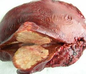
Multiple organ echinococcosis with involvement of the heart, liver and kidneys was diagnosed. The child was referred urgently to Amosov National Institute of Cardiovascular Surgery under National Academy of Medical Science of Ukraine (Kyiv), where the diagnosis was confirmed and the operation performed. Operatively: the pericardium was considerably thickened. The cyst was 6-8 cm in diameter, localised at the apex of the LV, with invasion of the entire ventricle wall, the cyst wall fibrosed. The contents of the cyst were aspirated using a syringe and an aspirator; the cyst was cut open; 5 cysts were removed from its cavity. The child was discharged in a satisfactory condition.
Thus, though cardiac is a very rare localisation, paediatricians and children cardiologists should keep it in mind, as there is a possibility of an effective surgical treatment.

