
Журнал «Здоровье ребенка» 1 (52) 2014
Вернуться к номеру
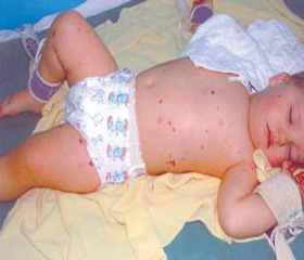
Case meningococcemia in a child is complicated by extensive necrosis of the skin
Авторы: Vodolazskaya G.R., Gavrish I.A. - State Institution "Institute of Emergency and Reconstructive Surgery of V.K. Husak" NAMS of Ukraine, Donetsk
Рубрики: Педиатрия/Неонатология
Разделы: Клинические исследования
Версия для печати
Introduction. Meningococcal disease is the leading cause of death from infections in early childhood (meningitis – in 15 %, sepsis – in 25 %, a combination of them – in 60 % of cases). Because of microcirculation and rheology disorders multiple necrotic areas form in different parts of the body. The most common skin lesions.
Materials and methods. We have the experience in the treatment of four children with necrotic lesions of the skin with meningococcemia. In this report a case of successfully treated a child with extensive necrosis of the skin was done.
Child G., 9 months, acutely ill 23.12.2012. During the five days he was treated in the isolation ward of the district hospital. 29.12.2012 it in a serious condition, with meningococcemia, impairment of consciousness (coma I grade) was transferred to the intensive care department of child infection of the central city hospital. 09.01.2013 the patient was transferred to the Donetsk burn center for surgical treatment.
Results and discussion. On admission the child's condition was extremely grave, he was in stupor and did not react to external stimuli. Respiratory failure occurred. Anemia, leukocytosis were expressed in the blood count.
Wounds were presented with black necrotic eschar, which was distributed to the upper and lower limbs, the trunk. Skin necrosis area was 40 % of the body surface. It was diagnosed sepsis and pneumonia. Overall severity of the condition is not possible to carry out surgical treatment. Intensive therapy (detoxification, antibacterial, transfusion of fresh frozen plasma, packed red blood cells, intravenous immunoglobulin G (5 % Bioven mono).
A week later, there was demarcated in the wounds, which allowed to perform necrectomy and ultrasonic cavitation of wounds. However, the regenerative processes in the wounds were sluggish current. In connection with this it was decided to refrain from autodermotransplantation. For health reasons, to reduce the endogenous intoxication and the elimination of the wound it was performed repeated debridement, ultrasonic cavitation wounds. Part of the wound was closed an autodermograft. On the other wounds allofibroblasts culture was transplanted.
Through the use of cellular technology we have been able to achieve active proliferation of fibroblasts in wounds that filled a deep wound defects with granulation tissue and get a high rate of engraftment autodermografts.
The child was discharged in satisfactory condition.
Conclusions. Timely surgery and intensive individual therapy can save the life of the child with meningococcemia, sepsis, widespread necrosis of the skin.
We consider it appropriate to continue to pursue surgical treatment of children with extensive necrotic lesions of the skin due to infectious diseases in specialized burn centers.
The burn centers should be equipped with modern technologies surgery and intensive care.
Throughout the period of surgical treatment is necessary supervision of a physician and pediatric intensive care.